Fungal folliculitis
Fungal folliculitis also called Malassezia folliculitis or Pityrosporum folliculitis, is an infection of hair follicles that typically manifests as a itch (pruritic), follicular papulopustular eruption distributed on the upper trunk of young to middle-aged adults caused by yeasts, specifically Malassezia furfur 1. The most common presentation is monomorphic papules and pustules, often on the chest, back, posterior arms, and face 2. Fungal folliculitis or Malassezia (Pityrosporum) folliculitis is commonly misdiagnosed as acne vulgaris (common acne) 3. Although often associated with common acne, fungal folliculitis may persist for years without complete resolution with typical acne medications.
Fungal folliculitis lesions are chronic (long term), erythematous (red), pruritic papules and pustules, which occur in a follicular pattern (related to the hair follicles). These lesions are usually present on the back and chest and occasionally, on the neck, shoulders, upper arms, and face. The diagnosis of fungal folliculitis or Malassezia folliculitis is based on clinical suspicion of the classic presentation of pruritic papulopustules found in a follicular pattern on the back, chest, upper arms, and, occasionally the neck. They are rarely present on the face. An improvement in the lesions with empiric anti-fungal therapy supports a clinical diagnosis of Pityrosporum folliculitis.
Malassezia furfur has been linked to several skin diseases, including seborrheic dermatitis, folliculitis, confluent and reticulated papillomatosis, and pityriasis versicolor 4.
Fungal folliculitis or Malassezia folliculitis is caused by overgrowth of yeast Malassezia species that are part of the normal inhabitant of human skin and not by exogenous species and only causes disease under specific conditions 5. With the advancement of technology, 7 species of Malassezia are recognized: Malassezia furfur, Malassezia pachydermatous, Malassezia sympodialis, Malassezia globosa, Malassezia obtusa, Malassezia restricta, and Malassezia slooffiae 6. Although different species may be involved, all species have the same clinical presentation 7. Although minor discrepancies occurred on the particular order of most to least common, the main species identified on lesional skin were Malassezia globosa, Malassezia restricta, and Malassezia sympodialis 8. Malassezia globosa was formerly known as Pityrosporum orbiculare and Malassezia restricta resembles Pityrosporum ovale 9. These particular species were not only identified as most common on lesional skin, but also nonlesional skin of the same patient as well as healthy controls 10. Durdu et al 7 in Turkey identified the most common species in lesional samples to be Malassezia globosa (69.4%) based on recombinant deoxyribonucleic acid (rDNA) analysis. This was followed by Malassezia sympodialis, Malassezia restricta, and Malassezia furfur in order of most to least common. Furthermore, the same species were again identified on lesional and nonlesional samples of the same patient in 72 percent of cases. These results point toward the hypothesis that Malassezia furfur indeed results from an overgrowth of the normal cutaneous flora and not an exogenous species 11. These studies were done in different geographical locations and thus it is possible that this resulted in differences in commonality of identified species 8.
Fungal folliculitis or Malassezia folliculitis was originally assumed to be caused by Pityrosporum ovale, thus leading to the name Pityrosporum folliculitis 12. In fact, Potter et al 13 confirmed the link of Pityrosporum orbiculare and Pityrosporum ovale, which make up Malassezia furfur, to this disease, which was confirmed by Back et al 14. Malassezia furfur is an oval, monopolar budding yeast 15. It is a polymorphic, lipophilic micro-organism with a thick, multilayered cell wall 16.
Malassezia furfur yeast is found in the stratum corneum and pilar folliculi where it uses its own lipases and phospholipases to hydrolyse triglycerides from sebum into free fatty acids for their own nutritive lipid source, thus leading to proliferation 17. It is an opportunistic organism, which changes from the saprophytic phase to the pathogenic mycelian phase under certain conditions, such as increased temperature, greasy skin, sweating and immunosuppression 18. Mokronosova et al 19 relate this to a change in the composition of fatty acids of the sebaceous gland due to an increase in androgen concentration.
Oral antifungals are the most effective treatment as it has proven much more effective than topical agent and result in rapid improvement. Fluconazole is used more commonly than itraconazole due to its superior side effect profile 20.
Topical agents (eg, selenium sulfide shampoo, econazole solution) may also be used but should be reserved for those unable to tolerate oral treatment 21.
Isotretinoin and photodynamic therapy (PDT) have been used with some success in small case series 20.
The association with acne vulgaris may require combinations of both antifungal and acne medications.
Figure 1. Fungal folliculitis
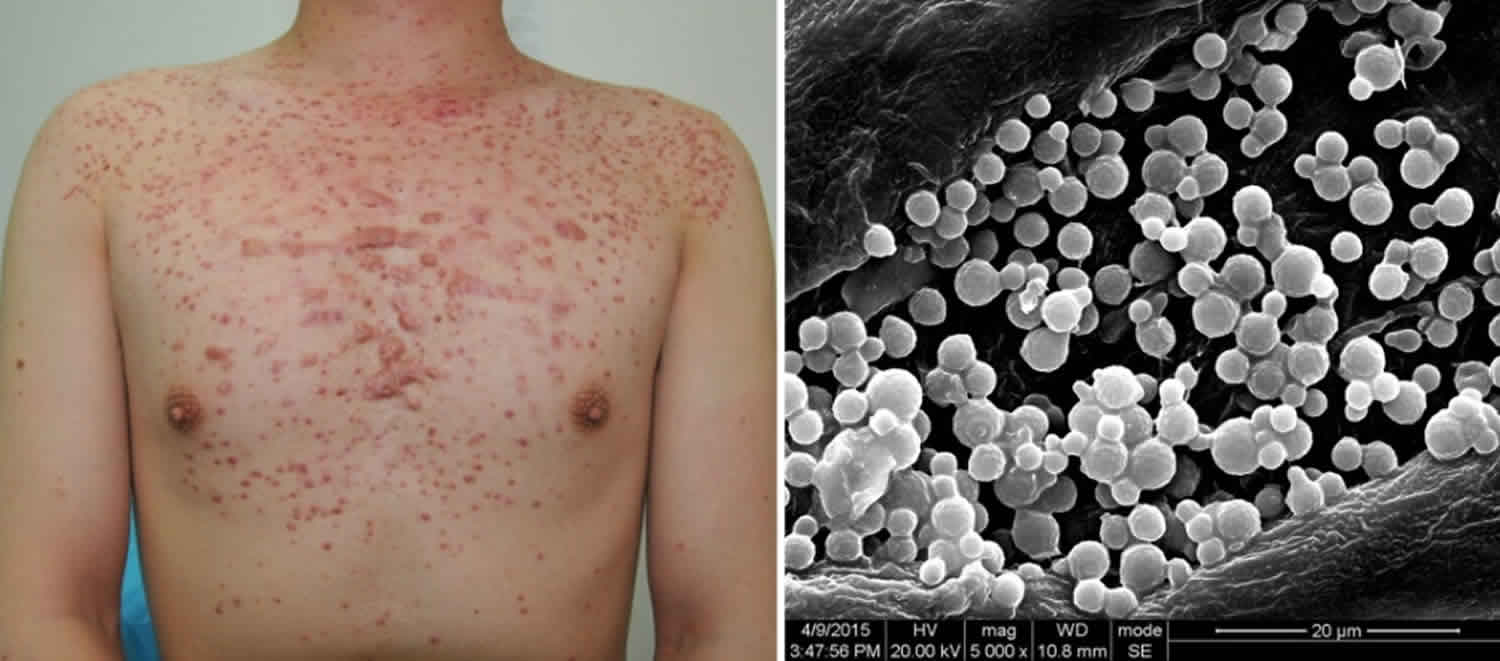
Footnote: A 25-year-old man with complaints of slightly pruritic, monomorphic follicular papules, pustules, and secondary keloid on the upper trunk and neck. Right: Scanning electron microscopy of the hair follicle from the upper trunk. This demonstrated a large number of globular or orbicular-ovate yeasts of budding daughter cell, with collar structure around the budding.
[Source 22 ]Figure 2. Facial fungal acne
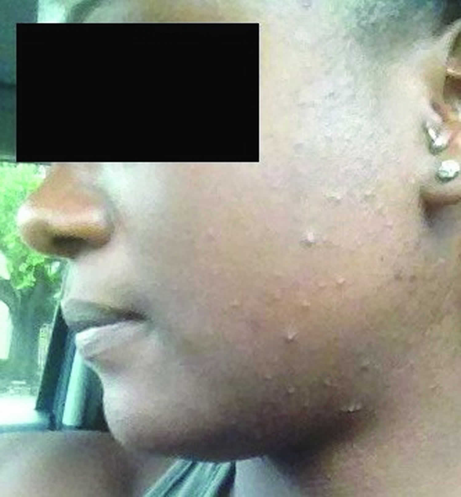
Footnote: Monomorphic papules and pustules on the cheek.
[Source 2 ]Figure 3. Fungal acne forehead
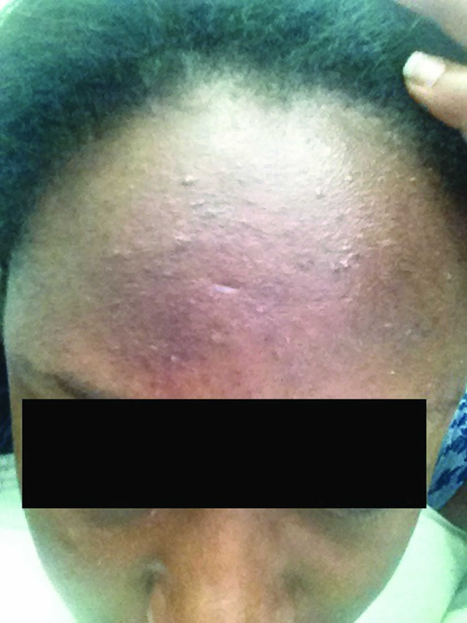
Footnote: Monomorphic papules and pustules on the forehead.
[Source 2 ]Fungal folliculitis causes
Fungal folliculitis or Malassezia folliculitis is caused by Malassezia yeasts, which are lipophilic. Several factors can lead to changes in immunity, sebum production, and the growth of skin flora. These factors help to produce favorable conditions for growth of these yeasts.
Systemic diseases and pharmacologic agents that encourage the growth of yeast, possibly because of alterations in immunity, include the following:
- Diabetes mellitus
- Cushing disease or Cushing syndrome
- Hodgkin’s disease 23
- Cancer treated with cetuximab (IMC-C225; marketed under the name Erbitux), a chimeric (mouse/human) monoclonal antibody epidermal growth factor receptor (EGFR) inhibitor for the treatment of metastatic colorectal cancer and head and neck cancer 24.
- HIV infection
- Corticosteroids and/or immunosuppressant therapy following organ transplantation 25.
- Crohn’s disease treated with infliximab a monoclonal antibody against tumor necrosis factor alpha 26.
An increase in sebum production, such as that in pregnancy 27 and high levels of androgens may potentiate the development of fungal folliculitis or Malassezia folliculitis.
Antibiotics can alter normal skin flora, allowing the yeast to proliferate.
Fungal folliculitis or Malassezia folliculitis more frequently occurs in environments of high heat and humidity.
Occlusion of the skin and hair follicles with cosmetics, lotions, sunscreens, emollients, olive oil, or clothing creates favorable conditions for fungal folliculitis or Malassezia folliculitis.
Anticonvulsant therapy and Down syndrome 28 are other conditions that are associated with fungal folliculitis or Malassezia folliculitis.
Other related and coexisting conditions may include the following:
- Seborrheic dermatitis
- Confluent and reticulated papillomatosis
- Systemic candidiasis 29
Some individuals seem to have an innate propensity for fungal folliculitis or Malassezia folliculitis. In one experiment, Malassezia yeasts were applied to occluded forearm skin in patients with fungal folliculitis or Malassezia folliculitis. Flares of fungal folliculitis or Malassezia folliculitis occurred at the application site. In the same experiment, fungal folliculitis or Malassezia folliculitis did not develop in patients with no prior diagnosis of the condition.
Fungal folliculitis pathophysiology
Malassezia furfur (ie, Pityrosporum ovale and Pityrosporum orbiculare) is a lipophilic, saprophytic, budding, unipolar, dimorphic, gram-positive, double-walled, oval-to-round yeast. Malassezia furfur is part of the normal skin flora. It is suggested that the similar yeasts Pityrosporum ovale and Pityrosporum orbiculare are actually identical and that they are morphologic variants of Malassezia furfur.
Malassezia yeasts are classified as superficial mycoses that by definition do not invade past the cornified epithelium. In fungal folliculitis or Malassezia folliculitis, however, the organism is present in the ostium and central and deep segments of the hair follicle.
Plugging of the follicle followed by an overgrowth of yeast that thrives in the sebaceous environment is believed to be the etiology. Malassezia yeasts require free fatty acids for survival. Usually, they are found in the stratum corneum and in pilar folliculi in areas with increased sebaceous gland activity such as the chest and back. The yeasts hydrolyze triglycerides into free fatty acids and create long-chain and medium-chain fatty acids from free fatty acids. The result is a cell-mediated response and activation of the alternative complement pathway, which leads to inflammation.
The inflammatory component of Malassezia furfur has many possible mechanisms. One possibility being Malassezia’s in vitro ability to induce keratinocyte production of inflammatory cytokines via Toll-like receptor 2 (TLR 2) 30. Among these inflammatory cytokines are interleukin (IL)-lα, IL-6, IL-8, IL-12, and tumor necrosis factor (TNF)-α along with anti-inflamatory cytokines IL-4 and IL-10 10. Malassezia activate complement cascades by both the classic and alternative pathways 16. Other possible mechanisms leading to inflammation include damage to the epithelial barrier function due to lipase and phospholipase activity of Malassezia, sensitization to cross-reactive allergens produced by Malassezia, and an irritant, nonimmunogenic stimulation of the immune system 30. This last mechanism is supported by the presence of an increased number of NK1+ and CD16+ cells within biopsies from lesional skin 31. While no difference was identified between the number of IL-associated cells between lesional and nonlesional skin, increased intensity was seen intercellularly in lesional skin.
Risk factors for developing fungal folliculitis
Other risk factors for developing fungal folliculitis include:
- High sebum production 2
- Hyperhidrosis (excessive sweating) 32
- Occlusion from emollients and sunscreens
- Antibiotic use 33
- Oral steroids such as prednisone (steroid acne)
- Immunosuppression 34.
- A history of hospitalization may also play a role in initial colonization 35.
Fungal folliculitis prevention
Fungal folliculitis recurrence is common, even after successful treatment 21.
Long-term prophylaxis with topical agents may be considered in those at high-risk or with multiple recurrences.
Periodic re-evaluation of predisposing factors is recommended.
Fungal folliculitis symptoms
Fungal folliculitis (Malassezia folliculitis) presents as intensely pruritic (itchy) as small 1 to 2mm uniform follicular itchy papules and pustules on the forehead, chin, neck, upper back, chest, shoulders and extensor aspect of the upper limbs 36. Fungal folliculitis (Malassezia folliculitis) is most commonly seen in adolescent and young adult males living in humid climates 2.
Durdu et al 7 found that 71.4 percent of fungal folliculitis or Malassezia folliculitis lesions in a study of 49 patients were found in more than one region. The most common location was the face (57.1%), followed by the back (53%), extensor surfaces of the arms (38.8%), chest (36.7%), and neck (18.3%). Furthermore, on the face, the chin and sides of the face were most commonly affected compared with the central facial lesions of acne vulgaris. Pruritus was a component of 79.6 percent of patients’ symptoms, and 10.2 percent even presented with excoriations.
Fungal folliculitis or Malassezia folliculitis is common in adolescents, likely due to increased sebaceous gland activity 37. Marcon et al 38 found that the frequency and density of colonization of the yeast is related to age and sebaceous gland activity. It is commonly found in people living in hot, humid climates, particularly those affected by excessive sweating, and is reported to be more common in males 7. Other predisposing factors include topical or oral antibiotic use, particularly tetracyclines, oral corticosteroid use, and immunosuppression 7. Malassezia furfur, made up of Pityrosporum orbiculare and ovale, have been detected in follicular contents of steroid acne 39. A study of 49 patients in Turkey, performed by Durdu et al 7, found the incidence of fungal folliculitis or Malassezia folliculitis to be four percent of patients attending their dermatology clinic, with an average age of 26 (range 12-62) years.
The main differential diagnoses of fungal folliculitis or Malassezia folliculitis are acne vulgaris; however, no comedones or cysts are associated with fungal folliculitis 40 and staphylococcal folliculitis. Often, patients have been treated with antibiotics or another medication appropriate for acne vulgaris, resulting in no improvement or worsening of their condition 41. Recalcitrant acne should be reevaluated for potential Pityrosporum infection 42.
Fungal folliculitis diagnosis
Clinical examination is usually sufficient for diagnosis. Laboratory investigations may be performed.
- Potassium hydroxide preparation of skin scrapings may reveal budding spores and hyphae 43.
- Other stains, including the May-Grunwald-Giema stain may also be helpful, but are less commonly used 44.
- Cultures are not routinely done, as malassezia species typically require special media for growth.
- High titers of circulating immunoglobulin G (IgG) antibodies against Pityrosporum ovale can also be detected in persons with fungal folliculitis 45.
Fungal folliculitis or Malassezia folliculitis is present on body locations in which Malassezia organisms are most abundant: back and chest, neck, shoulders, scalp, upper arms (occasional), and face 46. Under a Wood light, bright blue or white fluorescence is observed in clinically uninvolved follicles in the location of the lesions.
Dermoscopy may demonstrate perifollicular erythematous papules and pustules with “dirty-white” perilesional scale 47.
Fungal folliculitis (Malassezia folliculitis) may also be suspected by finding organisms within the hair follicles on histopathological examination of a skin biopsy.
Laboratory studies
A potassium hydroxide (KOH) preparation may be helpful for microscopic identification of the yeasts associated with fungal folliculitis or Malassezia folliculitis.
Culturing and identification of the organism are rarely performed, and the tests usually are not available. For Malassezia yeasts to grow, olive oil must be added to the culture media. This is not a routine study for the mycology laboratory.
Shibata et al 48 report the presence of galactomannan on cells of Malassezia species, which they suggest has not previously been reported in the literature. Results of their comparative antibody reaction studies lead them to suggest the potential for antigen detection as a diagnostic tool in Malassezia infection.
Fungal folliculitis treatment
It is important to address any predisposing factors at the outset, as fungal folliculitis or Malassezia folliculitis has a tendency to recur.
Both topical and oral antifungals are effective agents in the treatment of fungal folliculitis or Malassezia folliculitis. Oral antifungals have the advantage of dramatic, immediate clearing of the lesions and are the most effective treatment 49.
Patients have been successfully treated with oral pulse itraconazole and weekly fluconazole. Malassezia sympodialis is highly sensitive to terbinafine, while other species are more resistant to treatment with this medication 50. Many investigators have studied the efficacy of itraconazole, as this antifungal is excreted in high concentrations in sebum 35. Itraconazole is a broad-spectrum triazole, which is highly lipophilic and keratophilic with good oral absorption and extensive tissue distribution 51. Parsad et al 51 utilized 200mg itraconazole for seven days in 13 patients. Of these 13 patients, 11 showed negative mycological exam at week 5, compared to one in the placebo group, made up of 12 subjects. In one case report 41, 200mg itraconazole daily for two months resulted in nearly complete disappearance of lesions, promoting apparent cure; however, the patient did relapse after 12 months. Two weeks of 200mg itraconazole daily resulted in complete recovery of 79.6 percent of patients 7. Furthermore, itraconazole appears to delay relapses 52.
Many patients improve with topical azole medications, but some cases require oral therapy. A course of oral ketoconazole was previously the treatment of choice, but given the potential for severe adverse events with this medication, it is no longer recommended 53. Oral medication should be discontinued when the lesions resolve. Because relapse almost always occurs when treatment is withdrawn, topical ketoconazole is indefinitely continued after successful initial treatment with oral medication.
Adverse effects associated with oral antifungal medications include nausea, vomiting, diarrhea, abdominal pain, and hepatotoxicity; for this reason, some authors are proposing alternative treatments, including photodynamic therapy (PDT) 37. Other reasons for alternative therapies include infection relapses and possible drug resistance 54. Lee et al 55 did a pilot study using topical photodynamic therapy (PDT) with methyl aminolevulinate cream as a photosensitizer treatment for fungal folliculitis or Malassezia folliculitis. Patients underwent three sessions at two-week intervals with assessment at one month following the last treatment. Minimal side effects were noted, including a mild burning sensation after each treatment, which disappeared within 12 hours and slight hyperpigmentation, which disappeared within a couple of months. Out of the six patients included in this study, three presented with strong improvement, one with moderate improvement, one with mild improvement, and one with no improvement. However, this patient was an athlete and reported frequent sweating. The study reported no recurrence after four months. Proposed mechanisms for the effectiveness of this treatment include the complete destruction of fungal hyphae and inactivation of spores, which remain and survive on the skin following medical treatment, thus enabling recurrence, destruction of the pilosebaceous unit, and anti-inflammatory properties of red light, which influences cytokine release from macrophages 56.
Other topicals that are used to treat fungal folliculitis or Malassezia folliculitis are ciclopirox olamine cream, econazole cream, alcohol and salicylic acid solution (with or without benzoic acid 5%), propylene glycol 50% in water, and selenium sulfide shampoo 57. Other topical treatments with some reported success include tea tree oil, honey, tacrolimus, and cinnamic acid 58.
In cases associated with antibiotic use, discontinuing the antibiotic may be helpful.
Retinoids, which are used for comedones in acne, have no effect because no comedones are present in fungal folliculitis or Malassezia folliculitis 59.
Tetracycline does not help in fungal folliculitis or Malassezia folliculitis, and it may exacerbate the condition by further destroying the normal bacterial skin flora and allowing further spread of Malassezia yeasts.
Other studies suggest topical photodynamic therapy with methyl aminolevulinate may be a potential therapy for recalcitrant Malassezia folliculitis 60.
Fungal folliculitis prognosis
The prognosis in fungal folliculitis or Malassezia folliculitis is good. With treatment, fungal folliculitis or Malassezia folliculitis can completely resolve. Without treatment, fungal folliculitis or Malassezia folliculitis are classically pruritic.
Fungal folliculitis or Malassezia folliculitis may be a bothersome condition (ie, severe pruritus), but the lesions are benign. Some underlying conditions that predispose the patient to fungal folliculitis or Malassezia folliculitis include diabetes mellitus, immunodeficiency, and systemic candidiasis 61; these conditions may cause morbidity. Consider the presence of predisposing conditions when fungal folliculitis or Malassezia folliculitis is diagnosed.
References- Malassezia (Pityrosporum) Folliculitis. https://emedicine.medscape.com/article/1091037-overview
- Rubenstein RM, Malerich SA. Malassezia (pityrosporum) folliculitis. J Clin Aesthet Dermatol. 2014;7(3):37–41. https://www.ncbi.nlm.nih.gov/pmc/articles/PMC3970831
- Gaitanis G, Velegraki A, Mayser P, et al. Skin diseases associated with Malassezia yeasts: facts and controversies. Clin Dermatol. 2013;31:455–463.
- Gaitanis G, Velegraki A, Mayser P, Bassukas ID. Skin diseases associated with Malassezia yeasts: facts and controversies. Clin Dermatol. 2013 Jul-Aug. 31(4):455-63.
- Akaza N, Akamatsu H, Sasaki Y, Kishi M, Mizutani H, Sano A, Hirokawa K, Nakata S, Nishijima S, Matsunaga K. Malassezia folliculitis is caused by cutaneous resident Malassezia species. Med Mycol. 2009;47(6):618-24. doi: 10.1080/13693780802398026
- Gaitanis G, Magiatis P, Hantschke M, Bassukas ID, Velegraki A. The malassezia genus in skin and systemic diseases. Clin Microbiol Rev. 2012 Jan. 25(1):106-41.
- Durdu M, Guran M, Ilkit M. Epidemiological characteristics of Malassezia folliculitis and use of the May-Grunwald-Giemsa stain to diagnose the infection. Diagn Microbiol Infect Dis. 2013
- Ko JH, Lee YW, Choe YB, Ahn KJ. Epidemiologic study of Malassezia yeasts in patients with Malassezia folliculitis by 26S rDNA PCR-RFLP analysis. Ann Dermatol. 2011;23:177–184.
- Gupta AK, Batra R, Bluhm R, et al. Skin diseases associated with Malassezia species. J Am Acad Dermatol. 2004;51(5):785–798.
- Gaitanis G, Magiatis P, Hantschke M, et al. The Malassezia genus in skin and systemic diseases. Clin Microbiol Rev. 2012;25:106–141.
- Akaza N, Akamatsu H, Sasaki Y, et al. Malassezia folliculitis is caused by cutaneous resident Malassezia species. Med Mycol. 2009;47:618–624.
- Graham JH. Pityrosporurn ovale-in the discussion. Arch Dermatol. 1968;98:421.
- Potter BS, Burgoon CF, Johnson WC. Pityrosporum folliculitis. Report of seven cases and review of the Pityrosporum organism. Arch Dermatol. 1973;107:388–391.
- Back 0, Faergemann J, Hornqvist R. Pityrosporurm folliculitis: a common disease of the young and middle aged. J Am Acad Dermatol. 1985;12:56–61.
- Erchiga VC, Florencio VD. Malassezia species in skin diseases. Curr Opin Infect Dis. 2002;15:133–142.
- Ljubojevic S, Skerley M, Lipozencic J, et al. The role of Malassezia furfur in dermatology. Clin Dermatol. 2002;20:179–182.
- Nazzaro-Porro M, Passi S, Caprilli F, et al. Growth requirements and lipid metabolism of Pityrosporurn orbiculare. J Invest Dermatol. 1976;60:178–182.
- Roberts SOB. Pityriasis versicolor: a clinical and mycological investigation. Br J Dermatol. 1996;81:315–326.
- Mokronosova MA, Arzumanian VG, Gervazieva VB. Yeast-like fungi Malassezia (Pityrosporurn): clinical and immunological aspects of the study. Vestn Ross Akad Med Nauk. 1998;5:47–50.
- Hald M, Arendrup MC, Svejgaard EL, Lindskov R, Foged EK, Saunte DM; Danish Society of Dermatology. Evidence-based Danish guidelines for the treatment of Malassezia-related skin diseases. Acta Derm Venereol. 2015 Jan;95(1):12-9. doi:10.2340/00015555-1825.
- Abdel-Razek M, Fadaly G, Abdel-Raheim M, al-Morsy F. Pityrosporum (Malassezia) folliculitis in Saudi Arabia–diagnosis and therapeutic trials. Clin Exp Dermatol. 1995 Sep;20(5):406-9.
- https://commons.wikimedia.org/wiki/File:Pityrosporum_folliculitis_2.jpg
- Helm KF, Lookingbill DP. Pityrosporum folliculitis and severe pruritus in two patients with Hodgkin’s disease. Arch Dermatol. 1993 Mar. 129(3):380-1.
- Cholongitas E, Pipili C, Ioannidou D. Malassezia folliculitis presented as acneiform eruption after cetuximab administration. J Drugs Dermatol. 2009 Mar. 8(3):274-5.
- Blaes AH, Cavert WP, Morrison VA. Malassezia: is it a pulmonary pathogen in the stem cell transplant population?. Transpl Infect Dis. 2009 Aug. 11(4):313-7.
- Nasir A, El Bahesh E, Whitten C, Lawson A, Udall JN Jr. Pityrosporum folliculitis in a Crohn’s disease patient receiving infliximab. Inflamm Bowel Dis. 2010 Jan. 16(1):7-8.
- Parlak AH, Boran C, Topcuoglu MA. Pityrosporum folliculitis during pregnancy: a possible cause of pruritic folliculitis of pregnancy. J Am Acad Dermatol. 2005 Mar. 52(3 Pt 1):528-9.
- Kavanagh GM, Leeming JP, Marshman GM, Reynolds NJ, Burton JL. Folliculitis in Down’s syndrome. Br J Dermatol. 1993 Dec. 129(6):696-9.
- Klotz SA, Drutz DJ, Huppert M, Johnson JE. Pityrosporum folliculitis. Its potential for confusion with skin lesions of systemic candidiasis. Arch Intern Med. 1982 Nov. 142(12):2126-9.
- Baroni A, Orlando M, Donnarumma G, et al. Toll-like receptor 2 (TLR-2) mediates intracellular signaling in human keratinocytes in response to. Malassezia furfur. Arch Dermatol Res. 2006;297:280–288.
- Faergemann J, Bergbrant I-M, Dohse M, et al. Seborrhoeic dermatitis and Pityrosporurn (Malassezia) folliculitis:characterization of inflammatory cells and mediators in the skin by immunohistochemistry. Br J Dermatol. 2001;144:549–556.
- Gaitanis G, Velegraki A, Mayser P, Bassukas ID. Skin diseases associated with Malassezia yeasts: facts and controversies. Clin Dermatol. 2013 Jul-Aug;31(4):455-463. doi: 10.1016/j.clindermatol.2013.01.012
- Weary PE, Russell CM, Butler HK, Hsu YT. Acneform eruption resulting from antibiotic administration. Arch Dermatol 1969;100179-183.
- Rhie S, Turcios R, Buckley H, Suh B. Clinical features and treatment of Malassezia folliculitis with fluconazole in orthotopic heart transplant recipients. J Heart Lung Transplant. 2000 Feb;19(2):215-9.
- Faergemann J. In vitro and in vivo activities of ketoconazole and itraconazole against Pityrosporurm orbiculare. Antimicrob Agents Chemother. 1984;26:773–774.
- Poli F Differential diagnosis of facial acne on black skin. Int J Dermatol. 2012;51(Sl):24–26.
- Ayers K, Sweeney SM, Wiss K. Pityrosporum folliculitis: diagnosis and management in six female adolescents with acne vulgaris. Arch Pediatr Adolesc Med. 2005;159:64–67.
- Marcon MJ, Powell DA. Human infections due to. Malassezia spp. Clin Microbiol Rev. 1992;5:101–119.
- Katoh T, Irimajiri J. Pityriasis versicolor and Malassezia folliculitis. Nippon Ishinkin Gakkai Zasshi. 1999;40:69–71.
- Ayers K, Sweeney SM, Wiss K. Pityrosporum folliculitis: diagnosis and management in 6 female adolescents with acne vulgaris. Arch Pediatr Adolesc Med. 2005 Jan. 159(1):64-7.
- Marques SA, Pires de Camargo RM, Marques MEA, et al. Exuberant clinical presentation of probable Malassezia folliculitis in a young nonimmunosuppressed patient. An Bras Dermatol. 2012;87(3):459–462.
- Nakabayashi A, Sei Y, Guillot J. Identification of Malassezia species isolated from patients with seborrheic dermatitis, atopic dermatitis, pityriasis versicolor and normal subjects. Med Mycol. 2000;38:337–341.
- Thayikkannu AM, Kindo AJ, Veeraraghavan M. Malassezia – Can it be ignored? Indian J Dermatol. 2015;60(4):332-339.
- Durdu M, Güran M, Ilkit M. Epidemiological characteristics of Malassezia folliculitis and use of the May-Grünwald-Giemsa stain to diagnose the infection. Diagn Microbiol Infect Dis. 2013 Aug;76(4):450-7. doi: 10.1016/j.diagmicrobio.2013.04.011. Epub 2013 May 22.
- Faergemann J, Bergbrant IM, Dohse M, Scott A, Westgate G. Seborrhoeic dermatitis and Pityrosporum (Malassezia) folliculitis: characterization of inflammatory cells and mediators in the skin by immunohistochemistry. Br J Dermatol. 2001 Mar. 144(3):549-56.
- Aytimur D, Sengöz V. Malassezia folliculitis on the scalp of a 12-year-old healthy child. J Dermatol. 2004 Nov. 31(11):936-8.
- Jakhar D, Kaur I, Chaudhary R. Dermoscopy of pityrosporum folliculitis. J Am Acad Dermatol. 2019 Feb. 80 (2):e43-e44.
- Shibata N, Saitoh T, Tadokoro Y, Okawa Y. The cell wall galactomannan antigen from Malassezia furfur and Malassezia pachydermatis contains beta-1,6-linked linear galactofuranosyl residues and its detection has diagnostic potential. Microbiology. 2009 Oct. 155:3420-9.
- Hald M, Arendrup MC, Svejgaard EL, Lindskov R, Foged EK, Saunte DM. Evidence-based Danish Guidelines for the Treatment of Malassezia-related Skin Diseases. Acta Derm Venereol. 2014 Feb 20.
- Prohic A, Jovovic Sadikovic T, Krupalija-Fazlic M, Kuskunovic-Vlahovljak S. Malassezia species in healthy skin and in dermatological conditions. Int J Dermatol. 2015 Dec 29.
- Parsad D, Saini R, Negi KS. Short-term treatment of Pityrosporurm folliculitis: a double blind placebo-controlled study. J Eur Acad Dermatol Venereol. 1998;11(2):188–190.
- Caputo R, Barbareschi M. Itraconazole: new horizons. G Ital Dermatol Venereol. 2002;137:1–7.
- Prindaville B, Belazarian L, Levin NA, Wiss K. Pityrosporum folliculitis: A retrospective review of 110 cases. J Am Acad Dermatol. 2018 Mar. 78 (3):511-514.
- Lee JW, Kim BJ, Kim MN. Photodynamic therapy: new treatment for recalcitrant. Malassezia. folliculitis. Laser Surg Med. 2010 ;42: 192-196.
- Lee JW, Lee HI, Kim MN, et al. Topical photodynamic therapy with methyl aminolevulinate may be an alternative therapeutic option for the recalcitrant Malassezia folliculitis. Int J Dermatol. 2011;50:485–496.
- Horn M, Wolf P. Topical methyl aminolevulinate photodynamic therapy for the treatment of folliculitis. Photodermatol Photoimmunol Photomed. 2007;23:145–147.
- Viana de Andrade AC, Pithon MM, Oiticica OM. Pityrosporum folliculitis in an immunocompetent patient: clinical case description. Dermatol Online J. 2013 Aug 15. 19(8):19273.
- Pedrosa AF, Lisboa C, Gonçalves Rodrigues A. Malassezia infections: a medical conundrum. J Am Acad Dermatol. 2014 Jul. 71 (1):170-6.
- Goodfield MJ, Saihan EM. Failure of isotretinoin therapy in Pityrosporum folliculitis. J Am Acad Dermatol. 1988 Jan. 18(1 Pt 1):143-4.
- Lee JW, Lee HI, Kim MN, Kim BJ, Chun YJ, Kim D. Topical photodynamic therapy with methyl aminolevulinate may be an alternative therapeutic option for the recalcitrant Malassezia folliculitis. Int J Dermatol. 2011 Apr. 50(4):488-90.
- Gupta AK, Batra R, Bluhm R, Boekhout T, Dawson TL Jr. Skin diseases associated with Malassezia species. J Am Acad Dermatol. 2004 Nov. 51(5):785-98.


