Marjolin’s ulcer
Marjolin’s ulcer is the rare development of cutaneous squamous cell carcinoma (SCC) in the site of a pre-existing cicatrix or scar or ulcer 1. Marjolin ulcer is a cutaneous malignancy that arises in the setting of previously injured skin, longstanding scars, and chronic wounds. Marjolin’s ulcers are rare and are most commonly found in the lower extremity the legs and feet, especially the heel and plantar foot 2. The ulcers can also form on the head and neck 3. Marjolin ulcer most commonly forms at the site of an old thermal burn scar. However, Marjolin ulcers may also form from osteomyelitic lesions and, rarely, venous ulcers, pressure sores, surgical scars, animal bites, and vaccination scars 3.
Marjolin’s ulcers really reflect malignant degeneration arising within a pre-existing cicatrix or scar. In most instances, biopsied lesions demonstrate well-differentiated squamous cell tumors, although other epidermoid lesions are occasionally encountered 2.
Marjolin ulcers occur on average around 30 years after an injury to the skin that results in a scar or an ulcer (range 10–75 years) 4. Rarely, an acute Marjolin ulcer may develop between 6 weeks and 1 year of injury. It is estimated that around 2% of thermal burns scars and 0.7% of osteomyelitic lesions develop into Marjolin ulcers 5.
Marjolin ulcer can affect people of all age groups, sexes, and races. The average latency period from the time of the initial inciting wound to the discovery of malignant degeneration is between 30 and 35 years, with the average age at presentation being 59 years 6. Men are 2–3 times more likely be diagnosed with Marjolin ulcer than women, perhaps due to the increased frequency of burns in this population in general 7. Unlike other forms of skin cancer, all races and skin types can develop Marjolin ulcers 8.
Marjolin ulcer key points
- Marjolin ulcers most commonly arise from burn scars.
- The most common cell type identified is squamous cell carcinoma.
- All suspicious lesions should be biopsied.
- Confirmed cases of Marjolin ulcer should be excised with clear margins.
- Marjolin ulcers can be prevented with early excision and grafting of burn wounds.
- Prognosis is poor with a high rate of recurrence.
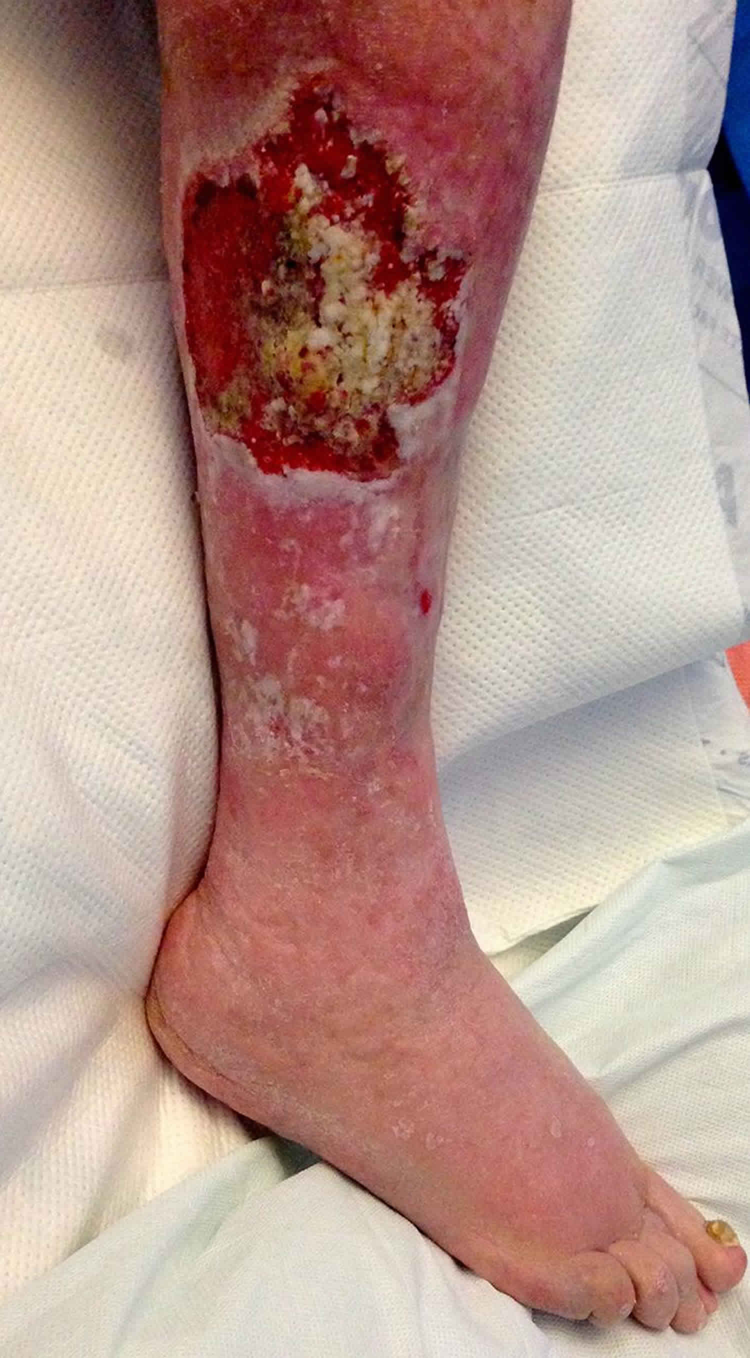
Figure 1. Marjolin ulcer
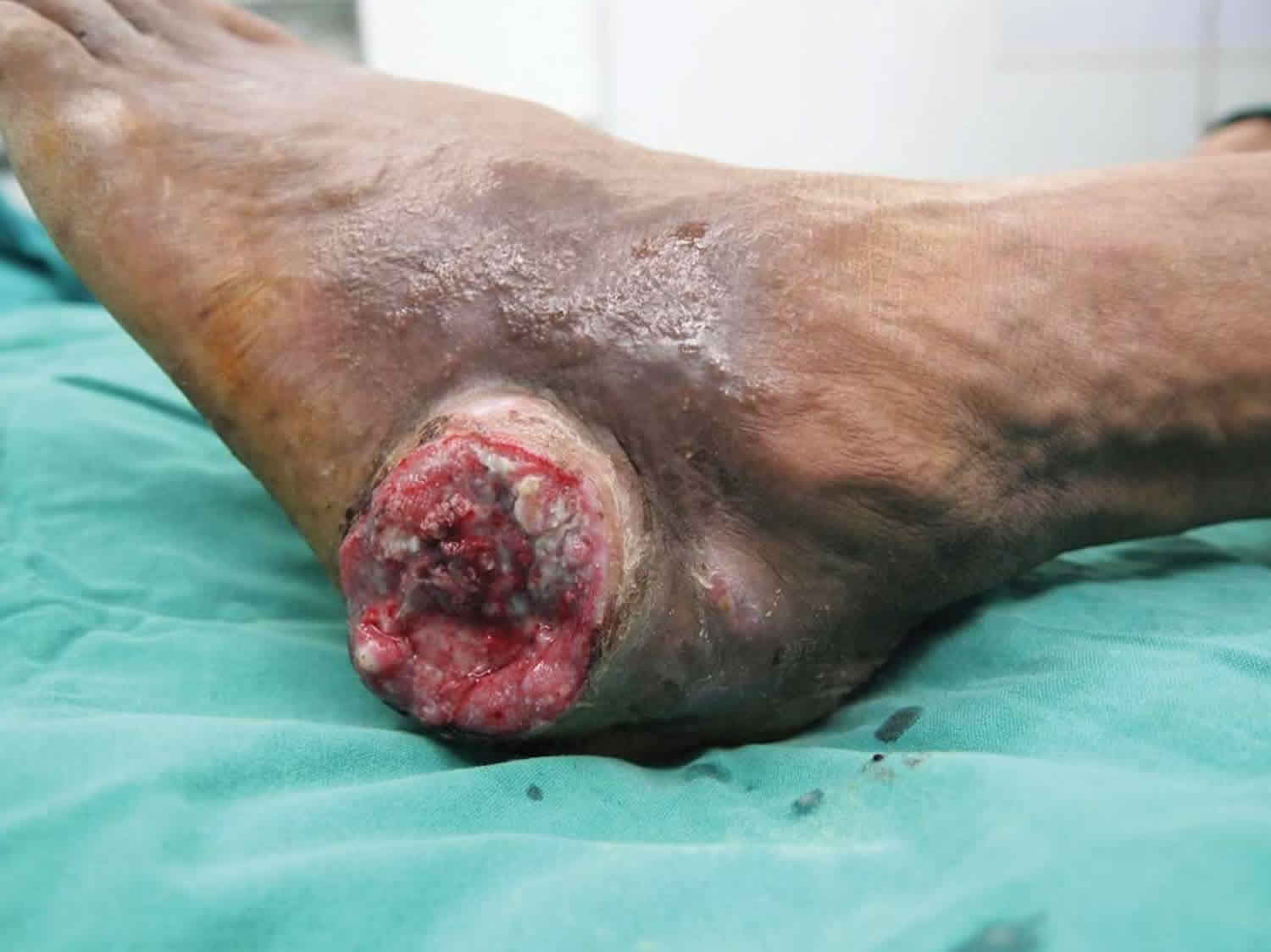
Footnote: Well-differentiated squamous cell carcinoma arising in an ulcer on the lateral aspect of the left ankle in a patient with simultaneous carcinomatous ulcers on the medial and lateral aspects of the ankle.
[Source 9 ]Figure 2. Marjolin ulcer radiology
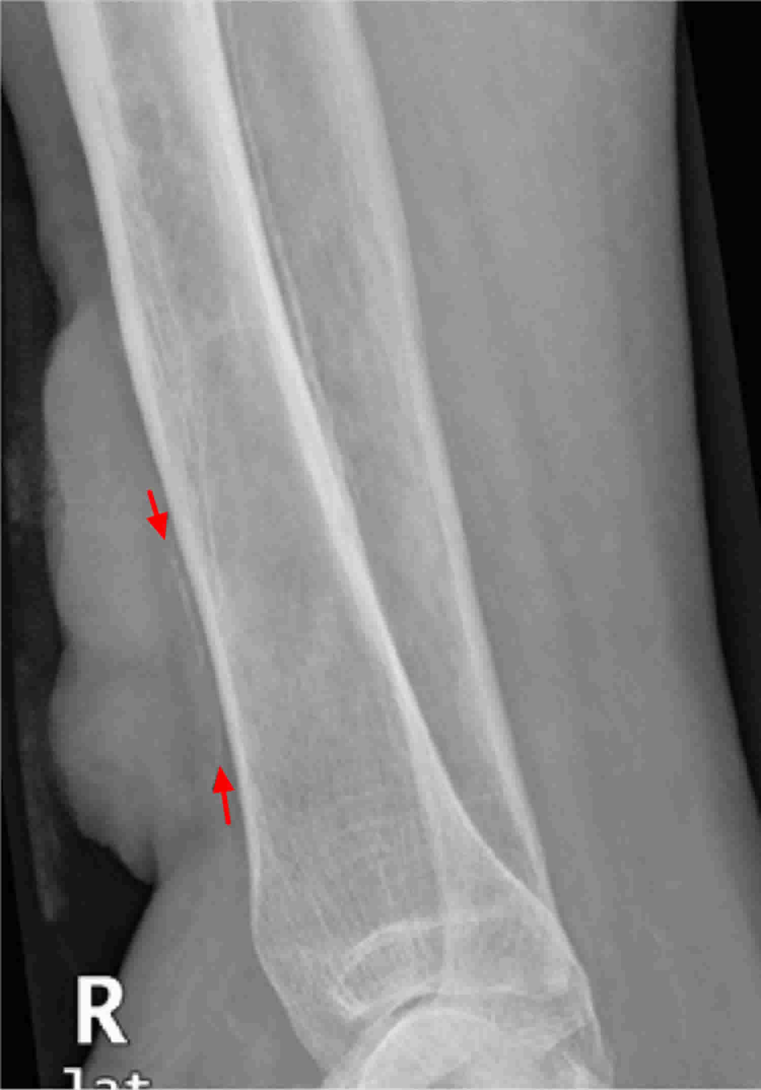
Footnote: A radiograph of right lower leg reveals a lobulated soft tissue mass over the anterolateral surface of the leg. The underlying tibia shows lamellated, thick periosteal reaction and an underlying ill defined lucent lesion in the medullary shaft.
[Source 10 ]Marjolin ulcer causes
The exact reason why Marjolin ulcers develop is unknown. Most theories suggest that injury and scar formation lead to destruction of the local blood and lymphatic vessels, making the area an immune-privileged site. This protects the scar from anti-tumor antibodies and permits the transformation and malignant degeneration of the skin. Chronic inflammation, irritation or trauma to the area are also thought to contribute to the process 4.
Burn scars are the most common inciting condition that leads to the development of Marjolin ulcers 11. Malignant degeneration occurs in 0.7% to 2.0% of burn scars that have been allowed to heal by secondary intention 12. Other chronic inflammatory etiologies that lead to Marjolin ulcers include traumatic wounds, venous stasis ulcers, osteomyelitis, pressure ulcers, radiation dermatitis, stings, bites, and hidradenitis suppurativa 13. Individuals that are immunocompromised, either due to disease state or medication, are at increased risk for malignant conversion 14.
Marjolin ulcer signs and symptoms
Marjolin ulcer usually presents as a nonhealing, ulcerative, or indurated lesion appearing in a chronic wound or scar 11. It may steadily increase in size, have excessive granulation tissue, rolled elevated margins, foul-smelling pus, bleed easily on contact, and be painful 15.
On examination, Marjolin ulcers are usually flat and indurated (hardened), with elevated margins. A less common, less aggressive type of Marjolin ulcer is slow-growing, and presents as an exophytic, papillary ulcer, which grows outward with finger-like projections 16.
Other clinical signs that suggest Marjolin ulcer formation include exophytic granulation tissue, bleeding, and regional lymphadenopathy 12. The ulcerative form is more common and more aggressive than the exophytic form. Superinfection of the wound may be the first presenting symptom. Lesions can occur anywhere but most frequently affect the lower extremities, followed by the scalp, upper extremities, torso, and face 17. Clinicians should pay close attention to all old scars on routine physical examinations and carefully note changes from prior visits.
Marjolin ulcer diagnosis
A Marjolin ulcer should be suspected when an ulcer persists for more than 3 months at the site of a scar 4.
Diagnosis is by incisional biopsy of suspicious areas of the ulcer. Cancerous change is detected on histological examination of the specimen. This may require a multidisciplinary approach, as Marjolin ulcer can be misdiagnosed as pseudoepitheliomatous hyperplasia 18.
Magnetic resonance imaging (MRI) may be performed to assess the degree of soft tissue and bone involvement 19.
Histopathology
Squamous cell carcinoma is the most frequent malignancy identified on histopathologic examination (80% to 90%) 20. Most lesions are well-differentiated, but poorly differentiated subtypes have also been identified. The second most common cell type is basal cell carcinoma (9.6%), followed by melanoma (2.4%). Rare cases of sarcoma, dermatofibrosarcoma, mucoepidermoid carcinoma, and leiomyosarcoma have been described 17.
Staging
No specific TNM (tumor, node, metastasis) staging criteria exist for Marjolin ulcers. After a biopsy has been performed, the histopathologic type of tumor can be used to stage the disease. For example, if the biopsy reveals squamous cell carcinoma, existing criteria established by the American Joint Committee on Cancer (AJCC) can be used 11. Marjolin ulcers are staged as other cutaneous squamous cell carcinomas, using the TNM (tumor, node, metastasis) classification system. This depends on the size of the lesion and how far it has metastasised to the lymph nodes and other organs.
Marjolin ulcer differential diagnoses
Differential diagnoses for Marjolin ulcer include ulcers or skin cancers resulting from other conditions. These include:
- Recurrent cutaneous squamous cell carcinoma at the site of a previously excised carcinoma
- Basal cell carcinoma or other cancer (eg, melanoma or sarcoma) at the site of a scar; these are sometimes called Marjolin ulcer.
- Diabetic foot ulcer — these are often painless and surrounded by thickened skin.
- Venous ulcer — these tend to be irregular and superficial, surrounded by hyperpigmentation due to capillary leakage.
- Arterial ulcer — these are more painful on elevation of the foot, with punched-out borders; the patient may have cramping pain on walking.
- Pyoderma gangrenosum — these start as a blister or pustule at the site of minor injury, and becomes painful, purulent and dusky-purple with an overhanging edge.
Marjolin ulcer treatment
Marjolin ulcer is usually treated by wide local excision 4. The most widely accepted treatment options include Mohs surgery, wide local excision with 1 to 2 cm margins, though this is uncommon due to the time, expense and expertise required 21. Patients with aggressive cancer may have sentinel lymph node biopsy. Lesions that involve the bone require amputation 22.
Mohs surgery can be considered in lesions on the face, scalp, hands, feet, areolae, and other areas where improved cosmesis is desired 23. Amputation is reserved for advanced-stage disease when wide local excision and Mohs surgery are not possible 24. Defect coverage with local flap, free flap, or avascular graft is often performed but remains controversial as some studies have shown an increased risk of recurrence with reconstruction 25. After resection, close follow-up is necessary due to the high risk of recurrence.
Lymph node dissection is controversial but can be considered with positive lymph nodes on physical or ultrasound examination 26. Adjuvant radiation and chemotherapy are indicated if surgical resection is not possible. Agents that have been suggested include topical 5-fluorouracil, methotrexate, l-phenylalanine, and platinum-based therapy 11. Radiotherapy is recommended with regional lymph node metastases, high-grade lesions (grade III), and lesions greater than 10 cm in diameter 27.
Radiotherapy and chemotherapy have not been shown to be effective in treating Marjolin ulcers. However, radiotherapy may be used for palliation in cases where surgery is not possible or refused 5.
Patients diagnosed with Marjolin ulcer require long-term follow-up, at least for 3 years 28.
Marjolin ulcer prognosis
Prognosis depends most strongly on whether the cancer has already spread to lymph nodes 29. Although most Marjolin ulcers are well-differentiated, lesions are aggressive and carry a poor prognosis. Metastases are found in up to 27% of patients compared to 3% for other causes of squamous cell carcinoma 17.
Overall 3 year survival for Marjolin ulcer is 65–75%, and 10 year survival is 34%. One-quarter of patients with ulcers present that have already metastasized, for whom, 3 year survival is 35–50% 4.
Recurrence after surgery is common, at a rate of 20–30% within 3 years 4.
High grade (grade II or III), the presence of nodal metastases, and lower extremity location indicate a worse prognosis 13.
References- Marjolin ulcer. https://dermnetnz.org/topics/marjolin-ulcer
- Pekarek B, Buck S, Osher L. A Comprehensive Review on Marjolin’s Ulcers: Diagnosis and Treatment. J Am Col Certif Wound Spec. 2011;3(3):60–64. doi:10.1016/j.jcws.2012.04.001 https://www.ncbi.nlm.nih.gov/pmc/articles/PMC3601857
- Kerr–Valentic MA, Samimi K, Rohlen BH, Agarwal JP, Rockwell WB. Marjolin’s ulcer: modern analysis of an ancient problem. Plast Reconstr Surg 2009; 123: 184–91.
- Pekarek B, Buck S, Osher L. A comprehensive review on Marjolin’s ulcers: diagnosis and treatment. J Am Col Certif Wound Spec 2011; 3(3): 60–4.
- Aydoğdu E, Yildirim S, Aköz T. Is surgery an effective and adequate treatment in advanced Marjolin’s ulcer?. Burns 2005; 31: 421–31. DOI: 10.1016/j.burns.2005.02.008
- Pekarek B, Buck S, Osher L. A Comprehensive Review on Marjolin’s Ulcers: Diagnosis and Treatment. J Am Col Certif Wound Spec. 2011 Sep;3(3):60-4.
- Cocchetto V, Magrin P, Paula RA, Aidé M, Razo LM, Pantaleão L. Squamous cell carcinoma in chronic wound: Marjolin ulcer. Dermatology Online Journal 2013; 19(2): 7
- Bradford PT. Skin cancer in skin of color. Dermatol Nurs. 2009 Jul-Aug;21(4):170-7, 206; quiz 178.
- Shen, R., Zhang, J., Zhang, F., Du, Y., Liang, W., Xu, L. … Chen, X. (2015). Clinical characteristics and therapeutic analysis of 51 patients with Marjolin’s ulcers. Experimental and Therapeutic Medicine, 10, 1364-1374. https://doi.org/10.3892/etm.2015.2699
- Sawhney S, Jain R, Kakaria A, Chopra P. Marjolin’s Ulcer: Radiographic and magnetic resonance appearances in two cases. Sultan Qaboos Univ Med J. 2009;9(2):162–166. https://www.ncbi.nlm.nih.gov/pmc/articles/PMC3074775
- Elkins-Williams ST, Marston WA, Hultman CS. Management of the Chronic Burn Wound. Clin Plast Surg. 2017 Jul;44(3):679-687.
- Saaiq M, Ashraf B. Marjolin’s ulcers in the post-burned lesions and scars. World J Clin Cases. 2014 Oct 16;2(10):507-14.
- Bazaliński D, Przybek-Mita J, Barańska B, Więch P. Marjolin’s ulcer in chronic wounds – review of available literature. Contemp Oncol (Pozn). 2017;21(3):197-202.
- Giesey R, Delost GR, Honaker J, Korman NJ. Metastatic squamous cell carcinoma in a patient treated with adalimumab for hidradenitis suppurativa. JAAD Case Rep. 2017 Nov;3(6):489-491.
- Choa R, Rayatt S, Mahtani K. Marjolin’s ulcer. BMJ 2015 Aug; 351: h3997. DOI: 10.1136/bmj.h3997
- Opara KO, Otene IC. Marjolin’s ulcers: a review. Nigerian Health Journal 2011; 11(4): 107–11.
- Sadegh Fazeli M, Lebaschi AH, Hajirostam M, Keramati MR. Marjolin’s ulcer: clinical and pathologic features of 83 cases and review of literature. Med J Islam Repub Iran. 2013 Nov;27(4):215-24.
- Bozkurt M, Kapi E, Kuvat SV, Ozekinci S. Current concepts in the management of Marjolin’s ulcers: outcomes from a standardized treatment protocol in 16 cases. J Burn Care Res 2010; 31(5): 776–80. DOI: 10.1097/BCR.0b013e3181eed210
- Chiang KH, Chou AS, Hsu YH, Lee SK, Lee CC, Yen PS, Ling CM, Lee WH, Lin CC, Chang PY et al. Marjolin’s ulcer: MR appearance. AJR Am J Roentgenol 2006; 186: 819–20. DOI: 10.2214/AJR.04.1921
- Elkins-Williams ST, Marston WA, Hultman CS. Management of the Chronic Burn Wound. Clin Plast Surg. 2017 Jul;44(3):679-687
- Rowe DE, Carroll RJ, Day CL. Prognostic factors for local recurrence, metastasis, and survival rates in squamous cell carcinoma of the skin, ear, and lip: implications for treatment modality selection. J Am Acad Dermatol 1992; 26: 976–90.
- Shah M, Crane JS. Marjolin Ulcer. [Updated 2020 Jan 2]. In: StatPearls [Internet]. Treasure Island (FL): StatPearls Publishing; 2020 Jan-. Available from: https://www.ncbi.nlm.nih.gov/books/NBK532861
- Iqbal FM, Sinha Y, Jaffe W. Marjolin’s ulcer: a rare entity with a call for early diagnosis. BMJ Case Rep. 2015 Jul 15;2015
- Challa VR, Deshmane V, Ashwatha Reddy MB. A Retrospective Study of Marjolin’s Ulcer Over an Eleven Year Period. J Cutan Aesthet Surg. 2014 Jul;7(3):155-9.
- Oruç M, Kankaya Y, Sungur N, Özer K, Işık VM, Ulusoy MG, Uysal A, Koçer U. Clinicopathological evaluation of Marjolin ulcers over two decades. Kaohsiung J. Med. Sci. 2017 Jul;33(7):327-333.
- Metwally IH, Roshdy A, Saleh SS, Ezzat M. Epidemiology and predictors of recurrence of Marjolin’s ulcer: experience from Mansoura Universityxs. Ann R Coll Surg Engl. 2017 Mar;99(3):245-249.
- Copcu E. Marjolin’s ulcer: a preventable complication of burns? Plast. Reconstr. Surg. 2009 Jul;124(1):156e-64e.
- Yu N, Long X, Lujan-Hernandez JR, et al. Marjolin’s ulcer: a preventable malignancy arising from scars. World J Surg Oncol 2013; 11: 313. DOI: 10.1186/1477–7819–11–313
- Ryan RF, Litwin MS, Krementz ET. A new concept in the management of Marjolin’s ulcers. Ann Surgery 1981; 193: 598–604.


