Hidrocystoma
Hidrocystoma also known as cystadenoma, Moll gland cyst, and sudoriferous cyst, is a rare, benign, cystic lesion of the skin – can be either eccrine or apocrine sweat glands, and are often found on the head and neck region 1. Hidrocystoma tends to grow slowly and usually occur on the face or scalp, commonly affecting the eyelid. The common solitary translucent eyelid cyst is an apocrine hidrocystoma. Multiple cysts on the lower eyelid are eccrine hidrocystomas. They affect both males and females, are more common in adults, and are usually asymptomatic. Furthermore, apocrine hidrocystomas are not affected by temperature whereas eccrine hidrocystomas can grow in size with heat exposure and grow smaller in cooler temperatures.
Eccrine hidrocystomas and apocrine hidrocystomas have a very similar presentation, and their distinction from other head and neck cyst-like lesions must ultimately be verified on biopsy and by careful examination under the microscope. For both types, cystic lesions such as epidermal inclusion cysts, mucoid cysts, hemangioma, and lymphangioma are considered in the differential. Attention is given to cysts (mostly apocrine hidrocystomas) that resemble basal cell carcinoma of the eyelid or malignant melanoma because of their color (typically blue-black). A biopsy of the lesions is very important to exclude these diagnoses.
Some patients may be uncomfortable with the appearance of hidrocystomas and may seek treatment for cosmetic purposes. These lesions may be removed by simple excision, cauterization, or with lasers such as the CO2 laser. Flattening of the lesions with the use of botulinum toxin A injections have also been reported.
Table 1. Differentiation between eccrine hidrocystoma and apocrine hidrocystoma
| Eccrine Hidrocystomas | Apocrine Hidrocystomas | |
|---|---|---|
| No. of lesions | Solitary or multiple | Primarily solitary (but occasionally manifesting as multiple lesions) |
| Color | Ranges from flesh-colored to light-blue tint | Ranges from dark-blue tint to black |
| Size | 1 to 6 mm in diameter | 3 to 15 mm in diameter |
| Age group affected | 30-75 years | 30-75 years |
| Gender distribution | More prevalent among females than among males | Females and males are affected in equal numbers |
| Epithelium types | 1-2 layers of cuboidal epithelium | Single/double cuboidal-columnar epithelium |
| Stains | S-100 positive (solitary type), periodic acid-Schiff negative | S-100 negative, periodic acid-Schiff positive |
| Special cellular features | No decapitation of cells, no secretory cells | Decapitation of secretory cells, papillary projections seen under microscope |
| Body location | Malar, periorbital, chest, axilla, neck | Face, ears, head, chest, shoulders |
| Possible treatments | Puncture and drainage, electrodessication, carbon dioxide laser, anticholinergic topical creams; placement in cold temperature | Puncture and drainage, electrodessication, carbon dioxide laser, anticholinergic topical creams |
| Special features | Worse in hot, humid weather | No change with temperature |
Eccrine hidrocystoma
Eccrine hidrocystomas are small and tense thin-walled cysts, ranging from 1 to 6 mm in diameter, and can occur as single or multiple lesions 2. They are found predominantly in adult females and are located mostly on the periorbital and malar regions.
Eccrine hidrocystomas are prevalent in adults between 30 and 70 years of age 2. Solitary eccrine hidrocystomas are equally prevalent among males and females; however, the multiple eccrine hidrocystomas are mainly seen in female patients 3.
Apocrine hidrocystoma
Apocrine hidrocystomas arise from the proliferation of apocrine glands, the glands of Moll along the eyelid margins and are usually solitary, with a diameter of 3 to 15 mm 2. Apocrine lesions are also found mostly on the head and neck and along the eyelid margin near the inner canthus 4. Apocrine lesions are less likely than eccrine lesions to occur at periorbital regions; nevertheless, the general distribution of lesions tends to occur in similar locations on the body for apocrine and eccrine hidrocystomas 2.
Apocrine hidrocystomas affect the same age groups as do eccrine hidrocystomas, and rarely occur during childhood or adolescence. They can appear as single or multiple cystic lesions. Although a significant number of cases of multiple apocrine hidrocystomas have been reported in the literature, the multiple type is rare in the general population 5.
Hidrocystoma causes
The exact cause of the hidrocystoma is unknown but it is hypothesized that the eccrine hidrocystoma is due to the obstruction of the eccrine sweat gland which causes the retention of secretions and the appearance of a dilated cystic structure. The apocrine hidrocystoma is believed to be an adenoma of the coil structure of the apocrine sweat gland. It is important to understand the two different types of sweat glands to distinguish between apocrine and eccrine hidrocystomas. Apocrine sweat glands are found only on certain areas of the body such as the eyelids, armpits, areolae, external ear, and the genital region. The ductal openings of the apocrine sweat gland are also more closely associated to the eyelashes compared to the eccrine sweat glands. Eccrine sweat glands are found distributed throughout the whole body.
Eccrine hidrocystomas usually result from dilation of cystic excretory eccrine glands due to retention of sweat and dilation or blockage of the sweat duct. Under the microscope, eccrine hidrocystomas appear as unilocular cysts, which usually contain a single cystic cavity composed of 1 or 2 layers of cuboidal cells. They are located within the mid-dermal to superficial layers of the skin, especially around the eyes. Unlike their apocrine counterpart, there are no secretory cells seen and decapitation of cells is not viewed under the microscope 2. Histochemically, they stain positive for S100 protein (solitary type), whereas apocrine hidrocystomas do not. The cyst and epidermis do not communicate, and the periodic acid-Schiff (PAS)-positive granules are not observed 2.
Apocrine hidrocystomas appear as unilocular or multilocular under the microscope. The cyst wall is composed of an inner layer of secretory columnar epithelium. The epithelium is either a single or double layer of cuboidal-columnar epithelium, which lies above an outer myoepithelial cell layer. Periodic acid-Schiff (PAS)- positive granules are observed in the presence of lipofuscin granules. Apocrine hidrocystomas also encompass papillary projections, which are not usually seen in the eccrine hidrocystomas. These projections are vascular connective tissue, covered by the secretory epithelium. Papillary projections then appear on the lumen of the cysts as an outgrowth extension from the wall of the cyst 4.
Associated syndromes
The inherited disorders that are most commonly associated with the presence of multiple eccrine/apocrine hidrocystomas are Goltz-Gorlin syndrome and Schopf-Schulz-Passarge syndrome.
Goltz-Gorlin also known as Jessner-Cole syndrome, or focal dermal hypoplasia tends to occur sporadically, with few familial cases having X-linked dominant transmission; it occurs mostly in females. Its cardinal features are microcephaly; midfacial hypoplasia; malformed ears; microphthalmia; periocular multiple hidrocystomas; papillomas of the lip, tongue, anus, and axilla; skeleton abnormalities; and mental retardation 6.
Schopf-Schulz-Passarge is an autosomal recessive syndrome characterized by multiple eyelid apocrine hidrocystoma, palmoplantar hyperkeratosis, hypodontia, and hypotrichosis. It is further characterized by hypotrichosis, cysts of the eyelids, and multiple periocular apocrine hidrocystomas 7.
Graves’ disease has also been associated with multiple eccrine hidrocystomas 8, possibly due to hyperhidrosis, which is seen in hyperthyroid patients. This is further supported by the disappearance of lesions after treatment of hyperthyroidism 8.
Hidrocystoma signs and symptoms
Eccrine hidrocystomas are benign tumors of sweat gland origin and are classified into 2 major groups: the Smith type, which is the most prevalent solitary type, and the Robinson, or multiple, type. They are typically dome-shaped, have an amber, brown, or bluish tint, and range from 1 to 6 mm in diameter. During hot or humid weather, these lesions will grow in size and/or multiply in number. Eccrine hidrocystomas usually do not involve the eyelid margin but rather are distributed around the eyelid skin; this, along with a lighter color presentation, typically differentiates it from the apocrine type 2.
The clinical presentation of apocrine hidrocystomas is very similar to that described above. They appear as dome-shaped, solitary, clear, cystic nodules with a smooth surface, and the color varies, ranging from flesh-colored to blue-black. Although the solitary hidrocystoma is the most common clinical presentation, multiple lesions on the face and neck have been reported 9. The cysts tend to stay asymptomatic and will follow a gradual course until a certain size is reached. There is no evidence of seasonal variations associated with apocrine hidrocystoma, whereas patients tend to present with eccrine hidrocystomas mostly during hot, humid weather 10.
Hidrocystoma diagnosis
Although hidrocystomas are benign lesions, a biopsy may be performed to make an accurate diagnosis to rule out other malignant tumors of the eye. Multiple hidrocystomas of the eyelid may also be associated with Schöpf-Schulz-Passarge syndrome.
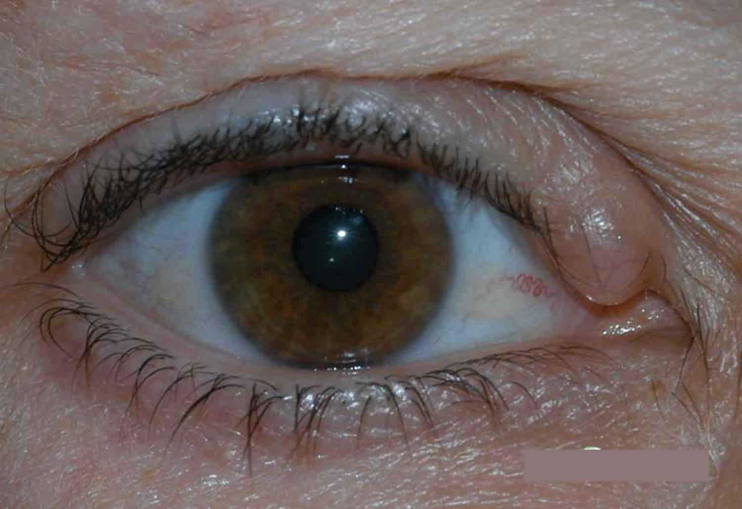
On physical examination, apocrine hidrocystomas are usually solitary, dome-shaped papules or nodules that range from flesh-colored to gray or blue. They range from a few millimeters to 1.5 cm and may involve the eyelid margin. In contrast, eccrine hidrocystomas are bluish or translucent-appearing papules that are usually located on the medial or lateral aspect of the eyelid that can be either solitary or multiple. They do not involve the eyelid margin but may appear close to this margin. They range from 1 mm to 5 mm in size.
Hidrocystoma treatment
The most common approach to the treatment of hidrocystomas (eccrine and apocrine) is simple needle puncture. However, in the Robinson type, a simple needle puncture will not produce lasting improvement. Multiple-type lesions have been successfully treated with topical 1% atropine or scopolamine creams, although anticholinergic side effects could cause patients to discontinue the treatment 11. Excision of hidrocystomas can lead to scars, and treatment involving incisions and drainage can lead to recurrence after 6 weeks; this can be overcome by cauterization and electrodessication of the cyst wall 2.
Gupta and colleagues 5 reported the use of electrodessication to be successful in the treatment of multiple apocrine tumors of less than 1 cm in diameter. Other methods that have shown success have been carbon dioxide laser vaporization and laser treatment 5. Tanzi and colleagues 12 effectively treated a patient with pulsed-dye treatment with a 585-nm laser, and no recurrence was observed after 18 months of treatment. Finally, avoiding hot temperatures or humid conditions will help prevent worsening of symptoms in patients diagnosed with eccrine-type hidrocystomas.
References- Sarabi K, Khachemoune A. Hidrocystomas–a brief review. MedGenMed. 2006;8(3):57. Published 2006 Sep 6. https://www.ncbi.nlm.nih.gov/pmc/articles/PMC1781304
- Alfadley A, Al Aboud K, Tulba A, Mazen M. Multiple eccrine hidrocystomas of the face. Int J Dermatol. 2001;40:125–129.
- Kaur C, Sarkar R, Kanwar AJ, Mohan H. Multiple eccrine hidrocystomas. J Eur Acad Dermatol Venereol. 2002;16:288–290.
- Alagheband M, Maida MF. Asymptomatic periorbital, bluish cystic papule. Cortlandt Forum. 2004;17:36–41.
- Gupta S, Handa U, Handa S, Mohan H. The efficacy of electrosurgery and excision in treating patients with multiple apocrine hidrocystomas. Dermatol Surg. 2001;27:382–384.
- Ascherman JA, Knowles SL, Troutman KC. Extensive facial clefting in a patient with Goltz syndrome: multidisciplinary treatment of a previously unreported association. Cleft Palate Craniofac J. 2002;39:469–473.
- Gira AK, Robertson D, Swerlick RA. Multiple eyelid cysts with palmoplantar hyperkeratosis–quiz case. Arch Dermatol. 2004;140:231–236.
- Kim YD, Lee EJ, Song MH, Suhr KB, Lee JH, Park JK. Multiple eccrine hidrocystomas associated with Graves’ disease. Int J Dermatol. 2002;41:295–297.
- del Pozo J, García-Silva J, Peña-Penabad C, Fonseca E. Multiple apocrine hidrocystomas: treatment with carbon dioxide laser vaporization. J Dermatolog Treat. 2001;12:97–100.
- Blugerman G, Schavelzon D, D’Angelo S. Multiple eccrine hidrocystomas: a new therapeutic option with botulinum toxin. Dermatol Surg. 2003;29:557–559.
- Armstrong DK, Walsh MY, Corbett JR. Multiple facial eccrine hidrocystomas: effective topical therapy with atropine. Br J Dermatol. 139:558–559. 1998 Sep.
- Tanzi E, Alster T. Pulsed dye laser treatment of multiple eccrine hidrocystomas: a novel approach. Dermatol Surg. 2001;27:898–900.


