What is an Id reaction
Id reaction also known as autoeczematisation, describes the occurrence of generalized eczema in response to a localized dermatosis or infectious and inflammatory skin conditions at a distant site 1. The very itchy rash that characterizes the Id reaction, which is considered immunologic in origin, has been referred to as dermatophytid 2, pediculid 3 or bacterid when associated with a corresponding infectious process 4. Clinical manifestations of the Id reaction are variable and depend on the cause of the eruption. Id reaction could be polymorphous including pompholyx-like reactions affecting hands and feet or more generalized papular eruptions. Histology of the id reaction often mimics that of the initially localized dermatosis or shows a spongiotic reaction pattern with varied intensity. Mild dermal edema and lymphocytic infiltration are reported.
Id reaction causes
The cause of Id reactions includes the following:
- Infections with dermatophytes, pulmonary histoplasmosis 5, mycobacteria 6, viruses 7, bacteria, or parasites (pediculosis) 3
- Contact dermatitis, stasis dermatitis, or other eczematous dermatoses 8
- Immunization reactions
- Papulonecrotic tuberculid 9 and some other tuberculids: These are now thought to be true cutaneous forms of tuberculosis and not id reactions because of the identification (by polymerase chain reaction) of Mycobacterium tuberculosis in lesions. However, id reactions have been reported following BCG vaccination 10.
While the exact cause of the Id reaction is unknown, the following factors are thought to be responsible:
- Abnormal immune recognition of autologous skin antigens,
- Increased stimulation of normal T cells by altered skin constituents 11,
- Lowering of the irritation threshold,
- Dissemination of infectious antigen with a secondary response,
- Hematogenous dissemination of cytokines from a primary site.
- Some cases have been related to medications and intravenous immune globulin 12.
Id reaction has also been noted with BCG therapy 10.
Id reaction signs and symptoms
Id reactions result from a variety of stimuli, including infectious entities and inflammatory skin conditions. Dermatological manifestations vary and depend on the etiology of the eruption. General history may include the following:
- Varying degrees of pruritus (itch) are typically noted.
- An acute onset of an extremely pruritic, erythematous, morbilliform, or papulovesicular eruption occurs 1-2 weeks after primary infection or dermatitis. Id reactions associated with stasis dermatitis are usually symmetrical and, in descending order of frequency, involve the forearms, thighs, legs, trunk, face, hands, neck, and feet.
- Id reactions are usually preceded by exacerbation of the preexisting dermatitis induced by infection, scratching, or inappropriate therapy. Id reaction to tinea incognito has been reported 13.
- Reactions have previously been reported after radiation treatment of tinea capitis.
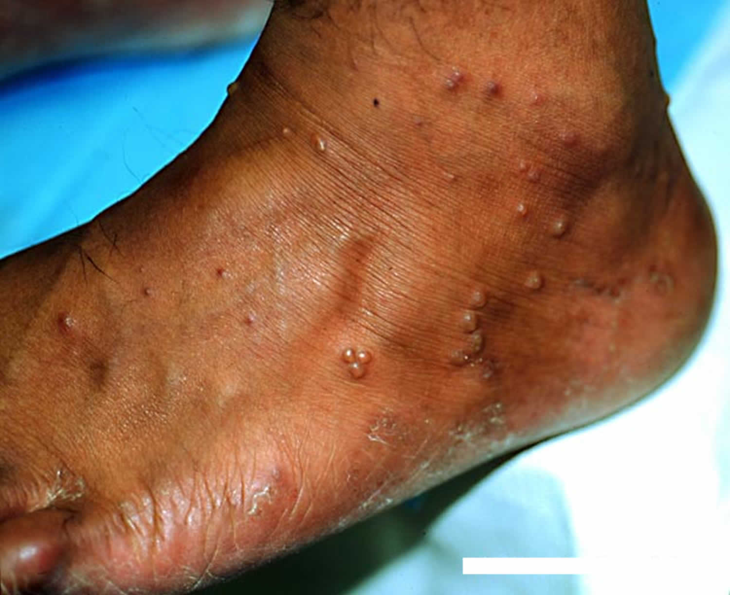
- Vesicles may be present on the hands or feet.
- Fingers may be tender.
- Travel history relating to infectious agent exposure may be relevant.
- A history of cultural or religious practices may indicate possible contact allergens leading to an Id reaction.
Id reaction complications
Id reaction complications can include secondary infection and secondary allergic contact dermatitis from topically applied medicaments/emollients.
Id reaction diagnosis
Clinical lesions of Id reactions are quite variable and are largely predicated on the inciting etiology. Lesions are, by definition, at a site distant from the primary infection or dermatitis. They are usually distributed symmetrically. Clinical forms include the following:
- A widespread, symmetrical eruption of small follicular papules associated with a kerion and a pompholyxlike eruption are usually associated with inflammatory tinea pedis (common) 14.
- An acute, intensely pruritic, symmetric maculopapular or papulovesicular reaction that involves the forearms, thighs, legs, trunk, face, hands, neck, and feet (in descending order of frequency) is typical of the Id reaction with stasis dermatitis (common).
- Erysipelas like eruption on the anterior leg secondary to a dermatophytosis may occur (less common).
- Extracutaneous manifestations include fever, anorexia, generalized adenopathy, splenomegaly, and leukocytosis (uncommon).
- The clinical picture may rarely mimic erythema multiforme 15.
Laboratory studies
Laboratory workup of Id reactions is clearly indicated for dermatophytids. Strict criteria include a proven dermatophyte infection and a positive skin test finding for a group-specific trichophytin antigen. Absence of fungi in the dermatophytid lesions and clearing of the dermatophytid after the fungus is eradicated are necessary to confirm a definitive diagnosis of a dermatophytId reaction.
Patch testing may be needed to exclude primary or secondary allergic contact dermatitis.
Skin biopsy
Biopsy for routine hematoxylin and eosin staining may be helpful in excluding noneczematous dermatoses, which may appear morphologically similar to an Id reaction.
Id reaction treatment
The goal is to adequately treat the underlying infection or dermatitis, which should lead to prompt resolution of the Id reaction. Recurrences are common, especially if the primary source is not treated adequately.
Treatment of the eruption includes the following:
- Systemic or topical corticosteroids. Corticosteroids help lesion resolution and provide symptomatic relief of pruritus. The strength and administration of a topical corticosteroid should be chosen based on the extent, location, and morphology of the eruption. Systemic corticosteroids may be used for severe or refractory eruptions.
- Prednisone (Orasone, Sterapred, Deltasone) is a commonly used oral agent. It is indicated for severe, prolonged, or anaphylactic reactions. It decreases late immune-mediated complications. Prednisone must be metabolized to the active metabolite prednisolone for effect. Conversion may be impaired in liver disease.
- Amcinonide (Cyclocort) suppresses mitotic activity and causes vasoconstriction. It stimulates the synthesis of enzymes needed to decrease inflammation.
- Fluocinonide (Fluonex, Lidex) is a high-potency steroid that inhibits cell proliferation. It is immunosuppressive, antiproliferative, and anti-inflammatory. It also has antipruritic and vasoconstrictive properties.
- Methylprednisolone (Depo-Medrol, Medrol, Adlone, Solu-Medrol) may decrease inflammation by reversing increased capillary permeability and suppressing polymorphonuclear neutrophils activity. It is indicated for severe, prolonged, or anaphylactic reactions. It decreases late immune-mediated complications.
- Wet compresses
- Systemic or topical antihistamines. Antihistamines relieve pruritus. Antihistamines may control itching by the blocking effects of endogenously released histamine.
- Diphenhydramine (Benadryl, Benylin, Caladryl, Dermapax) is a first-generation antihistamine with anticholinergic effects that binds to H1 receptors in the CNS and the body. Diphenhydramine is for symptomatic relief of symptoms caused by the release of histamine in allergic reactions. Diphenhydramine competitively blocks histamine from binding to H1 receptors. It has significant antimuscarinic activity and penetrates the CNS, which causes a pronounced tendency to induce sedation. Approximately half of those treated with conventional doses experience some degree of somnolence. A small percentage of children paradoxically respond to diphenhydramine with agitation.
- Loratadine (Claritin, Alavert) selectively inhibits peripheral histamine H1 receptors. It is tolerated well, with a rate of sedation not significantly different from placebo.
Id reaction prognosis
Id reaction prognosis is good once the inciting cause has been identified and appropriately treated. Morbidity results from symptoms of the Id reaction and the acute onset of the primary eruption.
References- Id Reaction (Autoeczematization). https://emedicine.medscape.com/article/1049760-overview
- Brown A, Sorey W. To Itch, Perchance to Scratch. Clin Pediatr (Phila). 2008 Nov 17.
- Brenner S, Ophir J, Krakowski A. Pediculid. An unusual -id reaction to pediculosis capitis. Dermatologica. 1984. 168(4):189-91.
- Chao SC, Lee YP, Lee JY. Eosinophilic cellulitis and panniculitis with generalized vesicular pustular id reaction after a molten aluminum burn. Dermatitis. 2010 Jun. 21(3):E11-5.
- Crum N, Hardaway C, Graham B. Development of an idlike reaction during treatment for acute pulmonary histoplasmosis: a new cutaneous manifestation in histoplasmosis. J Am Acad Dermatol. 2003 Feb. 48(2 Suppl):S5-6.
- Choudhri SH, Magro CM, Crowson AN, Nicolle LE. An Id reaction to Mycobacterium leprae: first documented case. Cutis. 1994 Oct. 54(4):282-6.
- Netchiporouk E, Cohen BA. Recognizing and managing eczematous id reactions to molluscum contagiosum virus in children. Pediatrics. 2012 Apr. 129(4):e1072-5.
- Trattner A, David M. Tefillin dermatitis. J Am Acad Dermatol. 2005 May. 52(5):831-3.
- Morrison JG, Fourie ED. The papulonecrotic tuberculide. From Arthus reaction to lupus vulgaris. Br J Dermatol. 1974 Sep. 91(3):263-70.
- Lowther C, Miedler JD, Cockerell CJ. Id-like reaction to BCG therapy for bladder cancer. Cutis. 2013 Mar. 91 (3):145-6, 151.
- Kasteler JS, Petersen MJ, Vance JE, Zone JJ. Circulating activated T lymphocytes in autoeczematization. Arch Dermatol. 1992 Jun. 128(6):795-8.
- Brazzelli V, Grassi S, Savasta S, Ruffinazzi G, Carugno A, Barbaccia V, et al. Pompholyx of the hands after intravenous immunoglobulin therapy for clinically isolated syndrome: a paediatric case. Int J Immunopathol Pharmacol. 2014 Jan-Mar. 27 (1):127-30.
- Al Aboud K, Al Hawsawi K, Alfadley A. Tinea incognito on the hand causing a facial dermatophytid reaction. Acta Derm Venereol. 2003. 83(1):59.
- Gorgievska-Sukarovska B, Skerlev M, Žele-Starčević L, Husar K, Halasz M. Kerion Celsi due to Microsporum canis with a Dermatophytid Reaction. Acta Dermatovenerol Croat. 2017 Jul. 25 (2):151-154.
- Atzori L, Pau M, Aste M. Erythema multiforme ID reaction in atypical dermatophytosis: a case report. J Eur Acad Dermatol Venereol. 2003 Nov. 17(6):699-701.


