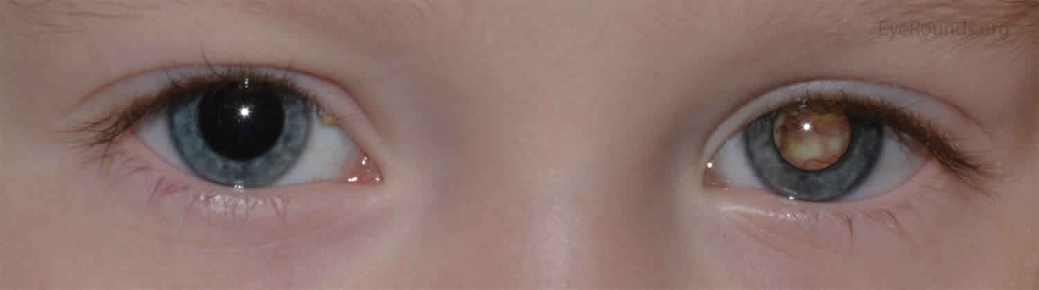
Leukocoria
Leukocoria also known as “white pupil,” is one of the primary signs of retinoblastoma 1. Leukocoria is present in over half of all infants presenting with retinoblastoma. Other conditions may also be associated with leukocoria as a presenting symptom, including: congenital malformations (e.g. persistent hyperplastic fetal vasculature); tumors (e.g. medulloepithelioma); vascular diseases (e.g. retinopathy of prematurity); inflammatory diseases (e.g. ocular toxocariasis); trauma (e.g. vitreous hemorrhage) 2.
Retinoblastoma is an uncommon type of malignancy occurring in 1 in 14000 to 1 in 20000 live births 3, but it is the most commonly encountered primary intraocular malignancy of childhood and accounts for 3% cases of all childhood cancers 4. Retinoblastoma is also the second most prevalent intraocular malignant tumor after uveal melanoma 5. In specialized care centers, survival rates are up to 95% with retention of vision in most cases, but it is lower in developing countries 5. Retinoblastoma is composed of retinoblasts (basophilic cells with hyperchromatic nuclei and scanty cytoplasm). Mostly retinoblastomas are undifferentiated, but different degrees of differentiation are present owing to the formation of structures known as rosettes. The tumor can be endophytic (in vitreous) and seeding of tumor cells throughout the eye, or it can be exophytic (in subretinal space), or it can demonstrate a mixed presentation. Optic nerve invasion can occur with the spread of tumor in subarachnoid space and into the brain. Metastatic spread occurs in regional lymph nodes, liver, lungs, bones, and brain 6.
Retinoblastoma occurs as a result of a mutation in the RB1 tumor suppressor gene located at the long arm of chromosome 13 at locus 14 (13q14) 7. Formation of tumor occurs when both the copies of the RB1 gene are mutated. In the case of bilateral retinoblastoma, there are 98% chances that the mutation is germline. Only 5% of cases of retinoblastoma have a family history. 95% of retinoblastoma cases are sporadic, of which 60 % of patients have unilateral disease with no associated germline mutation. Remaining patients present with germline mutations along with the development of multiple tumors.
- Heritable retinoblastoma: In this type of retinoblastoma, there is a mutation in one of the alleles of the RB1 gene in all body cells. When the second allele has a mutation as a result of some mutagenic event, it leads to the malignant transformation of cells. Due to the presence of the mutation in all cells, a large number of these children develop bilateral and multifocal retinoblastoma. Heritable disease patients are at significant risk of nonocular cancers such as pineoblastoma, osteosarcoma, soft tissue sarcomas, and melanomas: these malignancies usually occur in a particular age group 8. The chances of second malignancy are 6%, but the risk increases five-fold when external beam radiation has been used to treat the primary tumor.
- Non-heritable retinoblastoma: Non-heritable retinoblastomas are unilateral and are not transmitted. There is no risk of non-ocular cancers in these patients. In the case of unilateral retinoblastoma with no positive family history, it is non-heritable retinoblastoma, and the corresponding risk in each sibling and patient’s offspring is 1%. Almost 90% of unilateral retinoblastomas cases are of the nonhereditary form.
Leukocoria causes
Leukocoria is present in over half of all infants presenting with retinoblastoma.
Leukocoria differential diagnosis
Other conditions may also be associated with leukocoria as a presenting symptom, including 2:
- congenital malformations (e.g. persistent hyperplastic fetal vasculature);
- tumors (e.g. medulloepithelioma);
- vascular diseases (e.g. retinopathy of prematurity);
- inflammatory diseases (e.g. ocular toxocariasis);
- trauma (e.g. vitreous hemorrhage).
Leukocoria symptoms
Leukocoria also known as “white pupil,” is one of the primary signs of retinoblastoma 1. Leukocoria symptoms depend on the underlying cause of leukocoria.
Patients with retinoblastoma present mostly within the first year of age in the case of bilateral disease and within 3 years of age in case of unilateral disease. It is important to ask about family history of ocular malignancies. The most common presenting features are the following:
- Leucocoria: (whitish pupillary reflex): It is the most common presenting feature and accounts for 60% of cases.
- Strabismus: It is the second common presenting feature, and it is therefore important to perform fundus examination in all patients of childhood squint.
- Painful red eye: Painful red along with secondary glaucoma and associated buphthalmos can be present.
- Inflammation: Orbital inflammation resembling pre-septal or orbital cellulitis can also be a presenting feature.
- Visible extraocular growth
- Decreased vision
- Restriction of extraocular movements
- Metastatic disease: Metastatic disease involving lymph nodes, liver, lungs, brain, and bones is rare before ocular involvement.
Leukocoria diagnosis
Direct ophthalmoscopy
Red reflex testing with a direct ophthalmoscope is the simplest test, and leukocoria is easily observable. This method serves as a simple screening test 9.
Examination under anesthesia
Examination under anesthesia is necessary for measuring the corneal diameter, for tonometry, anterior chamber examination with a hand-held slit lamp, fundoscopy, cycloplegic refraction, and documenting all findings.
Ultrasound
To assess the size of the tumor, to observe calcifications, and it also helps to rule out similar conditions like coats disease.
Wide-Field Photography
Wide-field photography is used for analysis, documentation, and helps in the management of retinoblastoma.
CT scan
CT scans help in the detection of calcifications, but due to radiation risks, it is avoided upon making the primary diagnosis.
MRI
MRI is useful in the evaluation of optic nerve, extraocular extension, pineoblastoma, and to exclude similar diseases 10.
Systemic assessment
This includes physical examination, MRI orbit and brain, bone scan, bone marrow aspiration, and lumbar puncture.
Genetic studies
Genetic studies of blood samples and tumor tissue from patient and relatives 6.
Leukocoria treatment
Leukocoria treatment involves treating the underlying cause.
Treatment of retinoblastoma involves a multidisciplinary approach involving an ophthalmologist, pediatric oncologist, ocular pathologist, geneticist, allied health professional, and parents. Different treatment modalities employed in the treatment of retinoblastoma are;
Chemotherapy is the mainstay of treatment. It is also used in combination with local therapies. Intravenous carboplatin, etoposide, and vincristine are used in three to six cycles depending upon the grade of retinoblastoma. Single carboplatin or dual agent therapy can also be used and has shown favorable results in selective patients such as bridging therapy to avoid aggressive measures. Intravitreal melphalan is used in cases of vitreous seeding although it carries a small risk of extraocular dissemination. Chemoreduction is followed by cryotherapy or transpupillary thermal therapy to maximize tumor control.
Transpupillary thermal therapy is used mostly for focal consolidation after chemotherapy; however, it can be used as an isolated treatment. Transpupillary thermal therapy has a direct effect but also augments the effects of chemotherapy.
Cryotherapy the triple freeze-thaw technique is an option for pre equatorial tumors without deeper invasion or vitreous seedings.
Brachytherapy is used for an anterior tumor when there is no vitreous seeding and in cases of resistance to chemotherapy.
External beam radiotherapy is avoided when possible, especially in the case of heritable retinoblastoma because it can result in a second malignancy. Retinoblastomas are radiosensitive, but adverse effects include cataract, radiation neuropathy, radiation retinopathy, and hypoplasia of orbit 11.
Enucleation is performed when there is infiltration of the anterior chamber, neovascular glaucoma, invasion of the optic nerve, and if the tumor comprises more than half of the vitreous volume. It is also useful when chemotherapy has failed and in cases of diffuse retinoblastoma due to poor visual prognosis and a high risk of recurrence. Minimal manipulation should take place when performing enucleation, and a portion of the optic nerve of about 10 mm requires excision 12. Recent advances in enucleation techniques now allow the removal of a long segment of the optic nerve under direct vision 13.
Extraocular extension
Adjuvant chemotherapy for 6 months is given following enucleation when there is retrolaminar or massive choroidal spread. When the extension of the tumor is up to the cut end of the optic nerve at enucleation, or it is through the sclera, then external beam radiation is used.
Follow up
Careful follow-up at repeated intervals is required after treatment for early diagnosis of recurrence or development of new tumor, especially in patients with inherited disease.
Retinoblastoma prognosis
Patients with intraocular retinoblastoma, particularly those who have access to modern health care facilities, have an excellent prognosis and an overall survival rate of more than 95% in developed countries. The most critical risk factor associated with poor prognosis is extraocular extension either through the sclera or through the invasion of the optic nerve. Patients who survive bilateral retinoblastoma are at an increased risk of developing non-ocular malignancies later in life the latent period for the development of the second tumor is usually 9 months. External beam radiotherapy decreases the latent period and increases the risk of the second malignancy in the first 30 years of life. The most prevalent type of second malignancy is a sarcoma. The survival of patients who have developed sarcoma is less than 50%.
References- A Stepwise Approach to Leukocoria. https://www.aao.org/eyenet/article/stepwise-approach-to-leukocoria
- Balmer A, Munier F. 2007. Differential diagnosis of leukocoria and strabismus, first presenting signs of retinoblastoma. Clinical Ophthalmology. 1(4):431-439.
- Darwich R, Ghazawi FM, Rahme E, Alghazawi N, Burnier JV, Sasseville D, Burnier MN, Litvinov IV. Retinoblastoma Incidence Trends in Canada: A National Comprehensive Population-Based Study. J Pediatr Ophthalmol Strabismus. 2019 Mar 19;56(2):124-130.
- Alkatan HM, Al Marek F, Elkhamary S. Demographics of Pediatric Orbital Lesions: A Tertiary Eye Center Experience in Saudi Arabia. J Epidemiol Glob Health. 2019 Mar;9(1):3-10.
- Ishaq H, Patel BC. Cancer, Retinoblastoma. [Updated 2019 Aug 4]. In: StatPearls [Internet]. Treasure Island (FL): StatPearls Publishing; 2019 Jan-. Available from: https://www.ncbi.nlm.nih.gov/books/NBK545276
- Kletke SN, Feng ZX, Hazrati LN, Gallie BL, Soliman SE. Clinical Predictors at Diagnosis of Low-Risk Histopathology in Unilateral Advanced Retinoblastoma. Ophthalmology. 2019 Sep;126(9):1306-1314.
- Zahn J, Chan MP, Wang G, Patel RM, Andea AA, Bresler SC, Harms PW. Altered Rb, p16, and p53 expression is specific for porocarcinoma relative to poroma. J. Cutan. Pathol. 2019 Sep;46(9):659-664.
- House RJ, Hsu ST, Thomas AS, Finn AP, Toth CA, Materin MA, Vajzovic L. Vascular Findings in a Small Retinoblastoma Tumor Using OCT Angiography. Ophthalmol Retina. 2019 Feb;3(2):194-195.
- Shafiq A. Seeing red in young children: the importance of the red reflex. Br J Gen Pract. 2015 Apr;65(633):209-10.
- Jenkinson H. Retinoblastoma: diagnosis and management–the UK perspective. Arch. Dis. Child. 2015 Nov;100(11):1070-5.
- Elaraoud I, Ch’ng S, Karl D, Kalogeropoulos D, Chavan R, Sharma A. Management of retinal detachment in retinoblastoma with globe conserving treatment. J Curr Ophthalmol. 2019 Mar;31(1):43-48.
- Schefler AC, Kim RS. Recent advancements in the management of retinoblastoma and uveal melanoma. F1000Res. 2018;7
- Pelton RW, Patel BC. Superomedial lid crease approach to the medial intraconal space: a new technique for access to the optic nerve and central space. Ophthalmic Plast Reconstr Surg. 2001 Jul;17(4):241-53.


