Ophthalmia neonatorum
Ophthalmia neonatorum also known as neonatal conjunctivitis, is defined as conjunctival inflammation occurring within the first 30 days of life 1. Ophthalmia neonatorum may be aseptic or septic 2. Conjunctivitis is swelling or infection of the membrane that lines the eyelids and covers the white part of the eye. Neonatal conjunctivitisc omplications range from mild hyperemia and scant discharge to permanent scarring and blindness.
The three main causes of ophthalmia neonatorum or neonatal conjunctivitis include:
- Chemical: Classically, the most common cause of neonatal conjunctivitis due to use of post-delivery use of ophthalmic silver nitrate used in the prophylaxis of ocular gonococcal infections. However, the incidence of chemical conjunctivitis in the United States has significantly decreased since replacement of silver nitrate with erythromycin ointment
- Bacterial (Chlamydia trachomatis trachomatis most common). Bacterial cause of neonatal conjunctivitis include:
- Chlamydia trachomatis (most common)
- Neisseria gonorrhoeae. Neisseria gonorrhoeae is one of the most severe and feared causes of neonatal conjunctivitis, requiring prompt diagnosis and treatment.
- S. aureus
- Pseudomonas aeruginosa. Pseudomonas, although rare, may lead to potentially blinding complications such as rapid corneal ulceration and perforation.
- Streptococcus spp. (including Streptococcu haemolyticus, Streptococcu pneumonia)
- Other bacteria include Klebsiella, Proteus, Enterobacter, Serratia, and Eikenella corrodens 3
- Viral. Herpes simplex virus (HSV)
Prevalence of neonatal conjunctivitis has decreased significantly in developed countries since the abandonment of silver nitrate as topical prophylaxis. Current estimates of prevalence of neonatal conjunctivitis in developed countries are typically < 0.5%. However, a higher incidence of neonatal conjunctivitis is still found certain regions of the world, particularly in developing countries. A recent study found an estimated prevalence of 17% among nearly 1000 newborn infants in Pakistan 4. Indicidence of neonatal conjunctivitis remains high in Africa 5.
Aseptic neonatal conjunctivitis most often is a chemical conjunctivitis that is induced by silver nitrate solution, which is used at birth for Crede prophylaxis of infectious conjunctivitis. Chemical conjunctivitis is becoming less common owing to the use of erythromycin ointment or povidone iodide in place of silver nitrate solution for the prophylaxis of infectious conjunctivitis.
Bacterial and viral infections are major causes of septic neonatal conjunctivitis, with Chlamydia being the most common infectious agent. Infants may acquire these infective agents as they pass through the birth canal during the birth process 2.
The incidence of infectious ophthalmia neonatorum or neonatal conjunctivitis ranges from 1-2%, depending on the socioeconomic character of the area. Chlamydia is the most common infectious agent that causes ophthalmia neonatorum in the United States, where 2%-40% of neonatal conjunctivitis cases are caused by Chlamydia 6.
In contrast, the incidence of gonococcal ophthalmia neonatorum has been reduced dramatically and causes less than 1% of cases of neonatal conjunctivitis 7.
The conjunctiva (a thin translucent mucous membrane) can be divided into palpebral, bulbar, and fornical, based on the location. The conjunctiva contains nonkeratinizing, squamous epithelium and a thin, richly vascularized substantia propria containing lymphatic vessels and cells, such as lymphocytes, plasma cells, mast cells, and macrophages. The conjunctiva also has accessory lacrimal glands and goblet cells.
The pathology of neonatal conjunctivitis is influenced by the anatomy of the conjunctival tissues in the newborn. The inflammation of the conjunctiva may cause blood vessel dilation, potentially dramatic chemosis, and excessive secretion. This infection tends to be more serious in neonates owing to their lack of immunity, lack of lymphoid tissue in the conjunctiva, and absence of tears at birth.
Ophthalmia neonatorum causes
The cause of ophthalmia neonatorum or neonatal conjunctivitis can be chemical, bacterial (Chlamydia trachomatis most common) or viral. Although several noninfectious and infectious agents can inflame the conjunctiva, the most common causes of neonatal conjunctivitis are silver nitrate solution and chlamydial, gonococcal, staphylococcal, and herpetic infections 8.
Silver nitrate solution
Crede’s method of instilling a drop of 2% aqueous solution of silver nitrate into a newborn’s eyes was first published in 1881 and significantly advanced the prevention of neonatal conjunctivitis 9.
Silver nitrate is a surface-active chemical that facilitates agglutination and inactivation of gonococci. Ironically, silver nitrate was later found to be toxic to the conjunctiva, particularly in higher concentrations, potentially causing a sterile neonatal conjunctivitis.
Chlamydial conjunctivitis
Chlamydia trachomatis is an obligate intracellular parasite and has been identified as the most common infectious cause of neonatal conjunctivitis 10.
The reservoir of the organism is the maternal cervix or urethra. Infants who are born to infected mothers are at high risk (approximately 25%-50%) of developing an infection 6. Chlamydial pneumonitis may also accompany neonatal conjunctivitis.
Neisserial conjunctivitis
Neisseria gonorrhoeae is a gram-negative diplococcus and is potentially the most dangerous and virulent infectious cause of neonatal conjunctivitis. As with chlamydia, maternal cervical and urethral mucosa provide a reservoir for N gonorrhoeae, which is acquired during birth.
Gonococci can penetrate intact epithelial cells and divide rapidly inside them. Diagnostic Gram or Giemsa stain smears obtained from genitourinary or ocular mucosal scrapings reveal characteristic gram-negative intracellular diplococci.
Gonococcal conjunctivitis must be absolutely excluded in every case of neonatal conjunctivitis to prevent potentially blinding corneal and conjunctival complications.
Other bacteria
The most commonly identified gram-positive organisms include Staphylococcus aureus, Streptococcus pneumoniae, Streptococcus viridans, and Staphylococcus epidermidis. These bacteria make up 30-50% of all cases of infectious neonatal conjunctivitis 11.
Gram-negative organisms, such as Escherichia coli, Klebsiella pneumoniae, Serratia marcescens, and Proteus, Enterobacter, and Pseudomonas species, also have been implicated. There has been one reported case of Eikenella corrodens neonatal conjunctivitis 12.
Infants of low birth weight and low gestational age with clinical signs of conjunctivitis in the neonatal intensive care unit (NICU) should be evaluated and treated for a gram-negative etiology 13.
Herpes simplex
Herpes simplex virus (HSV) is a rare cause of neonatal keratoconjunctivitis, found in less than 1% of cases 11 and can be associated with a generalized herpes simplex infection.
Most infants with such an infection acquire the disease during the birth process. Caesarean delivery is strongly considered when active maternal genital disease is recognized at term since the risk of transmitting HSV to the neonate during vaginal delivery is 25-60% 14.
Risk factors for developing ophthalmia neonatorum
Risk factors of neonatal conjunctivitis may include:
- Maternal infections harbored in the mother’s birth canal
- HIV-infected mothers 15
- Exposure of the infant to infectious organisms
- Increased birth weight 15
- Inadequacy of ocular prophylaxis immediately after birth
- Premature rupture of membranes (PROM) 16
- Ocular trauma during delivery
- Mechanical ventilation
- Prematurity
- Poor prenatal care
- Poor hygienic delivery conditions
- Post-delivery infection due to direct contact with health care workers or by aerosolization
- Silver nitrate exposure
Ophthalmia neonatorum prevention
Parents or care providers need to wash their hands frequently to prevent transmission of neonatal conjunctivitis. Prevention through good prenatal care and treatment of chlamydial, gonococcal, or herpetic infections during pregnancy remains the best preventative method. Pregnant women need to understand the importance of regular examinations to detect and treat sexually transmitted infections (STIs) such as herpes simplex, gonorrhea, and chlamydia in order to decrease the incidence of neonatal conjunctivitis.
- Chlamydial infections occur in 4–10% of pregnant women in the United States
- Infants whose mothers have untreated chlamydial infections have a 30–40% chance of developing conjunctivitis (incidence of 6.2 per 1000 live births)
Ophthalmia neonatorum prophylaxis
According to the 2012 Red Book, topical 0.5% erythromycin and 1% tetracycline are considered equally effective for prophylaxis of ocular gonorrhea infection in newborn infants 17. Each is available in single-dose tubes. Topical silver nitrate, povidone-iodine, and erythromycin are all effective in the prevention of nongonococcal nonchlamydial neonatal conjunctivitis. There is no agent that is currently effective in preventing the transmission of Chlamydia trachomatis from mother to baby 18. This is a change from the 2009 Red Book which stated that erythromycin or silver nitrate could prevent vertical transmission 7.
Povidone-iodine solution (2.5%) is effective in preventing neonatal ophthalmia. Povidone-iodine is widely in Europe. It is approved by the US Food and Drug Administration (FDA), but it is not commercially available in this country 18. Recent studies showed that significantly fewer chlamydial infections occurred with povidone-iodine than with silver nitrate or erythromycin (5.5 versus 10.5 and 7.4 percent, respectively). Neomycin and chlorimphenicol are additional topical prophylactic options.
Silver nitrate appears to be the best agent in areas where the incidence of penicillinase-producing Neisseria gonorrhoeae is significant 19.
The recommendations in the 2012 Redbook are for 2 drops of 1% silver nitrate or a 1 cm ribbon of antibiotic ointment (either erythromycin or tetracycline) placed into the lower conjunctival sac; both acceptable regimens for the prophylaxis of neonatal conjunctivitis 11. Erythromycin ointment is considered the best regimen for prophylaxis against neonatal conjunctivitis because of its efficacy against gonococcal and nongonococcal nonchlamydial pathogens and owing to its low incidence of causing a chemical conjunctivitis 19.
Systemic prophylaxis
Infants with possible infectious exposure in utero or during birth process should receive appropriate prophylaxis following birth in attempt to prevent ocular and systemic complications. Gonoccal prophylaxis includes single injection of ceftriaxone 50 mg/kg IM or IV in those neonates born to mothers with untreated or suspected gonococcal infection.
Other preventative measures include proper hand-washing techniques by peripartum and nursery staff.
Ophthalmia neonatorum symptoms
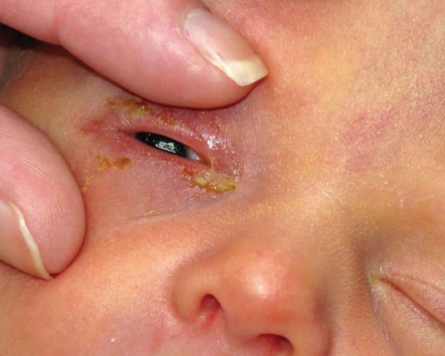
Presentations for different organisms may vary. Non-specific signs of neonatal conjunctivitis include conjunctival injection, tearing, mucopurulent or non-purulent discharge, swelling or edema of the conjunctiva (chemosis) and eyelid swelling. Typical findings may include redness (erythema) and swelling (edema) of the eyelids and palpebral conjunctiva (chemosis) and/or purulent eye discharge during the external eye exam. A Gram stain conjunctival smear should be performed in all cases.
Incubation period
Chemical conjunctivitis secondary to silver nitrate solution application usually occurs in the first day of life, disappearing spontaneously within 2-4 days.
Gonococcal conjunctivitis tends to occur 2-7 days after birth but can present later 11.
The onset of chlamydial conjunctivitis is usually later than gonococcal conjunctivitis; the incubation period is 5-14 days.
The expected incubation period for other nongonococcal, nonchlamydial conjunctivitis is also 5-14 days 11.
Herpetic conjunctivitis usually occurs within the first 2 weeks after birth and has an incubation period of approximately 6-14 days 11.
Gonococcal conjunctivitis
Gonococcal conjunctivitis tends to be the most serious form of ophthalmia neonatorum. Gonococcal conjunctivitis presents with the most rapid onset, usually occurring 24-48 hours following birth. Typically, patients develop a hyperacute conjunctivitis, associated with marked lid edema, chemosis, and purulent discharge. The classic presentation is severe bilateral purulent conjunctivitis.
Corneal ulceration may occur, particularly in the periphery, where massive limbal conjunctival chemosis traps inflammatory mediators and organisms, with rapid progression to perforation the cornea if treatment is delayed and endophthalmitis.
Patients also may have systemic manifestations, including rhinitis, stomatitis, arthritis, meningitis, anorectal infection, and septicemia.
Chlamydial conjunctivitis
The presentation of chlamydial conjunctivitis may range from mild hyperemia with scant mucoid discharge to eyelid swelling, chemosis, and pseudomembrane formation. Patients typically present with unilateral or bilateral watery discharge, which may become more copious and purulent later.
Although most cases are mild and self-limited, chlamydial conjunctivitis occasionally may be severe. Pseudomembranes, thickened palpebral conjunctiva, significant peripheral pannus, and corneal opacification may be present.
Blindness, although rare and much slower to develop than in gonococcal conjunctivitis, is generally not due to corneal involvement as in gonococcal conjunctivitis. Instead, eyelid scarring and corneal pannus can gradually progress to central corneal opacification by mechanisms reminiscent of trachoma.
A follicular reaction does not occur, because newborns have no requisite lymphoid tissue present in the conjunctiva.
Like gonococcal conjunctivitis, chlamydial conjunctivitis also may be associated with extraocular involvement, including pneumonitis, otitis, and pharyngeal and rectal colonization.
Neonatal conjunctivitis due to other agents
Neonatal conjunctivitis due to other microbial agents is usually milder.
Herpes simplex keratoconjunctivitis often presents in infants with generalized herpes simplex infections, characterized by corneal epithelial involvement or vesicles on the periocular skin. Serious systemic complications, such as encephalitis, may also occur in these neonates owing to their poor immunologic response.
Herpetic conjunctivitis
This type of neonatal conjunctivitis typically occurs within the first 2 weeks after birth.
Ocular involvement may follow systemic or central nervous system herpes infection, as well as vesicular lesions on the skin or lid margins.
Patients may present with nonspecific lid edema, moderate conjunctival injection, and a nonpurulent and often serosanguineous discharge, which may be unilateral or bilateral.
Microdendrites or geographic ulcers, rather than typical dendrites as seen in adults, are the most typical signs of herpetic keratitis in newborns.
Conjunctival membrane may be present.
Other bacterial conjunctivitis
Various organisms, including gram-positive and gram-negative bacteria, have been identified in neonatal conjunctivitis.
Classic clinical pictures are lid edema, conjunctival injection, chemosis, and discharge, which are variable and often indistinguishable from signs of other etiologies.
Although rarely implicated in neonatal conjunctivitis, Pseudomonas can lead to devastating consequences, such as rapid progression to corneal ulceration and perforation. If left untreated, Pseudomonas keratitis even can lead to endophthalmitis and subsequent death.
Chemical conjunctivitis
The clinical picture of chemical conjunctivitis is mild with transient tearing and conjunctival injection, spontaneously resolving within 2-4 days.
If the 1% silver nitrate used for neonatal conjunctivitis is provided in a large bottle, the solution can evaporate or settle, thereby becoming more concentrated over time. More concentrated silver nitrate solution may result in more severe responses, including, lid edema, chemosis, exudate, membranes or pseudomembranes, and permanent cicatricial damage to the conjunctiva or the cornea. This problem is obviated by using sealed, single-use ampules. Chemical conjunctivitis is becoming less common because of the substitution of alternative agents such as erythromycin ointment, tetracycline ointment, or povidone iodide in place of silver nitrate.
Ophthalmia neonatorum complications
Ocular complications of neonatal conjunctivitis include pseudomembrane formation, corneal edema, thickened palpebral conjunctivia, peripheral pannus formation, corneal opacification, staphyloma, corneal perforation, endophthalmitis, loss of eye, and blindness. Risk of complications can minimized with prompt diagnosis and appropriate antibiotic therapy.
If untreated, peripheral corneal ulceration may occur in Neisseria gonorrhoeae infection and rapidly progress to corneal perforation. Complications of gonococcal conjunctivitis and subsquent systemic involvement include arthritis, meningitis, anorectal infection, septicemia, and death.
When unrecognized and not immediately treated, Pseudomonas infection may lead to endophthalmitis and subsequent death.
Systemic complications of chlamydia conjunctivitis include pneumonitis, otitis, and pharyngeal and rectal colonization. Pneumonia has been reported in 10-20% of infants with chlamydial conjunctivitis 19.
HSV keratoconjunctivitis can cause corneal scarring and ulceration. Additionally, disseminated HSV infection often includes central nervous system involvement 14.
Ophthalmia neonatorum diagnosis
History
Time frame of signs/symptoms following birth play an important role in determining the most likely etiology and subsequent proper diagnosis and treatment:
- Chemical conjunctivitis (Typically presents within first 24 hours following birth)
- Neisseria gonorrhea (3-5 days after birth)
- Chlamydia trachomatis (5-14 days)
- HSV (1-2 weeks)
Physical examination
Your baby’s doctor will perform an eye exam on the baby. A thorough examination of the globe and periocular structures of a neonate suspected to have neonatal conjunctivitis is crucial. Corneal involvement should be investigated closely with and without fluorescein and blue cobalt light. A complete systemic examination should be performed by trained physician familiar with the physical exam of a neonate.
If the eye does not appear normal, the following tests may be done:
- Culture of the drainage from the eye to look for bacteria or viruses
- Slit-lamp exam to look for damage to the surface of the eyeball
Laboratory studies for neonatal conjunctivitis should include the following:
- Conjunctival scraping for Gram stain or Giemsa stain
- Conjunctival scraping for polymerase chain reaction (PCR) assay to detect chlamydia and gonorrhea
- Culture on chocolate agar and/or Thayer-Martin for N gonorrhoeae
- Culture on blood agar for other bacteria
- Culture of corneal epithelial cells for HSV if cornea is involved; PCR should also be considered in cases of possible HSV conjunctivitis
Culture and histology
Bacterial cultures on blood and chocolate agar are indicated in every case of neonatal conjunctivitis and remain the criterion standard despite newer diagnostic methods.
Since Chlamydia bacteria are obligate intracellular organisms, the culture specimens need to contain epithelial cells and not just exudative material. PCR is generally accepted as the most useful test for chlamydial conjunctivitis owing to its high sensitivity 19.
In cases in which gonorrhea is suspected, the agar should be inoculated immediately since Ngonorrhoeae is very sensitive to moisture and temperature changes.
Laboratory evaluation for the presence of HSV infection is indicated if a corneal epithelial defect is present, if vesicles are present on the eyelids or other parts of the body, and if the diagnosis cannot be made on ocular examination. The presence of HSV in tissue culture remains the criterion standard in the diagnosis of HSV, despite a high false-negative rate. HSV infections may be more rapidly diagnosed with PCR, and PCR testing for HSV is more sensitive than viral culture 19. Laboratory evaluation for suspected HSV becomes more important in neonatal disease because the clinical presentation may be highly atypical in an immunologically immature newborn.
Cytologic findings for various forms of conjunctivitis are as follows:
- Chemical conjunctivitis – Neutrophils, occasional lymphocytes on Gram stain
- Bacterial conjunctivitis – Bacteria, neutrophils on Gram stain
- Gonococcal conjunctivitis – Neutrophils, Gram-negative intracellular diplococci on Gram stain
- Chlamydial conjunctivitis – Neutrophils, lymphocytes, plasma cells on Gram stain; basophilic intracytoplasmic inclusions in epithelial cells on Giemsa stain
- Herpetic conjunctivitis – Lymphocytes, plasma cells, multinucleate giant cells on Gram stain; eosinophilic intranuclear inclusions in epithelial cells on Papanicolaou smear, but with low sensitivity
Newer diagnostic techniques
Nucleic acid amplification tests such as polymerase chain reaction (PCR) and transcription-mediated amplification are more sensitive than culture in detecting chlamydial and gonorrheal organisms 7.
PCR assays may have a higher sensitivity and similar specificity in diagnosing neonatal chlamydial conjunctivitis, compared with conventional methods 20.
PCR for HSV from conjunctival scrapings has high sensitivity and specificity, but it is expensive, not always readily available, and is usually reserved for the diagnosis of encephalitis. Direct florescent antibody (DFA) studies are useful for rapid detection, have high sensitivity and specificity, and can be used to type the virus 14.
Ophthalmia neonatorum treatment
Specific treatment is available for each cause of neonatal conjunctivitis. Preliminary presumptive treatment pending culture confirmation should be based on the clinical picture and the findings on Gram, Giemsa, and Papanicolaou stains.
Prior to birth, consider the risk of transmission of chlamydial, gonococcal, herpetic, and streptococcal pathogens to the fetus during vaginal delivery. Obtain cervical cultures if indicated and manage appropriately, including the possibility of a Caesarian delivery.
To confirm the presence of a sexually transmitted disease in the neonate, examine and treat the mother and her sexual partner(s). If necessary, therapy can be modified when the results of culture and sensitivity are known.
The treatment prior to laboratory results should include topical erythromycin ointment and an IV or IM third-generation cephalosporin. Prompt treatment of gonococcal conjunctivitis is important, since this organism can penetrate an intact corneal epithelium and rapidly cause corneal ulceration. Because of the rapid progression of gonococcal conjunctivitis, patients with acute neonatal conjunctivitis should be treated for gonococcal conjunctivitis until culture results are available; the treatment is altered according to the laboratory results.
In cases of chlamydial conjunctivitis, systemic treatment is necessary because of the significant risk for life-threatening pneumonia.
Infants with a potentially sexually transmitted disease, such as gonorrhea or chlamydia, should undergo evaluation for other sexually transmitted diseases, such as syphilis and HIV 21, as should the mother and her sexual partner(s).
Newborns with conjunctivitis are at risk for secondary infections, such as pneumonia, meningitis, and septicemia, which can lead to sepsis and death and thus should be admitted for full workup and treatment.
Bacterial conjunctivitis rarely fails to respond to treatment.
A consultation can be made with a pediatrician or pediatric infectious specialist in neonatal conjunctivitis, and the patient should be seen daily until response to treatment is confirmed.
Discharged patients should continue the treatment, according to clinical presentations and available culture results. Treatment may be modified later per culture results.
Avoid eye patching
Treatment of neonatal chemical conjunctivitis is not necessary. Lubrication with artificial tear preparations may ease mild discomfort.
Chemical conjunctivitis
No treatment required; supportive care only (may use artificial tears four times daily)
Typically disappears spontaneously within 2-4 days.
Neonatal Chlamydial conjunctivitis
This infection is treated with erythromycin drops four times daily plus oral erythromycin (50 mg/kg/day divided four times daily) for 2 to 3 weeks. While outpatient treatment is an option, hospitalization may be required. Evaluation of systemic involvement needed.
Topical treatment alone is ineffective. Topical erythromycin ointment may be beneficial as an adjunctive therapy.
Since the efficacy of systemic erythromycin therapy is approximately 80%, a second course sometimes is required.
Systemic treatment is important in cases of chlamydial conjunctivitis since topical therapy is ineffective in eradicating the bacteria in the nasopharynx of the infant, which could cause a life-threatening pneumonia if left untreated.
Neonatal gonococcal conjunctivitis
Topical irrigation with normal saline to remove mucopurulent discharge. Ceftriaxone in a single dose (25-50 mg/kg IM or IV, up to a maximum of 125 mg). If there is systemic disease, treatment is required for 7 to 14 days depending on the nature of the invasive infection. Bacitracin or erythromycin ointment every 2 to 4 hours. Hospitalization and evaluation for disseminated N. gonorrhea infection. Topical saline drops to remove discharge. Topical atropine if corneal involvement.
- Note: All neonates with gonococcal conjunctivitis should also be treated for chalmydia. Mother and sexual partner should be treated as well.
Other bacterial ophthalmia neonatorum
- Gram positive -Bacitracin ointment four times daily for 2 weeks
- Gram negative -Gentamicin, tobramycin or ciprofloxacin four times daily for 2 weeks
Neonatal herpetic conjunctivitis
Neonates with a suspected herpes simplex infection should be treated with systemic acyclovir to reduce the risk of a systemic infection.
An effective dose is Acyclovir 60 mg/kg/day IV divided three times daily plus vidarabine 3% ointment. The recommended minimal duration is 14 days, but a course as long as 21 days may be required.
Infants with neonatal HSV keratitis should also receive a topical ophthalmic drug, most commonly 1% trifluridine drops or 3% vidarabine ointment 5x/day for 14-21 days depending on presence or absence of CNS involvement. Topical ganciclovir 0.15% gel is now also available, although none of these topical agents is specifically approved for neonatal use 22.
Topical antibiotics can also be considered to prevent secondary bacterial infections in cases with significant epithelial defects.
Medical follow up
- Patients with neonatal conjunctivitis should be followed daily for signs of improvement or worsening, especially acutely due to concerns of rapidly progressing infectious complications such as those mentioned above.
- Patient should be followed closely by pediatrician for evaluation and treatment of potential systemic infection.
Ophthalmia neonatorum prognosis
Ophthalmia neonatorum or neonatal conjunctivitis usually responds to appropriate treatment, and the prognosis generally is good as long as early diagnosis is made and prompt medical therapy is initiated.
Antibiotics have significantly altered the prognosis of neonatal conjunctivitis, especially with Neisseria gonorrhoeae infection.
Mortality associated with neonatal conjunctivitis is due to systemic involvement of the infectious agent. No published information is available on mortality. Morbidity and mortality increases in cases of systemic involvement requiring hospitalization and intensive monitoring.
References- Neonatal Conjunctivitis. https://eyewiki.org/Neonatal_Conjunctivitis
- Neonatal Conjunctivitis (Ophthalmia Neonatorum). https://emedicine.medscape.com/article/1192190-overview
- Chhabra MS, Motley WW 3rd, Mortensen JE. Eikenella corrodens as a causative agent for neonatal conjunctivitis.J AAPOS. 2008 Oct;12(5):524-5.
- Gul SS, Jamal M, Khan N. Ophthalmia neonatorum. J Coll Physicians Surg Pak. 2010 Sep;20(9):595-8.
- Isenberg SJ et al. A double application approach to ophthalmia neonatorum prophylaxis. Br J Ophthalmol. 2003 Dec;87(12):1449-52.
- American Academy of Pediatrics. Chlamydia Trachomatis. Pickering LK, Baker CJ, Kimberlin DW, Long SS, eds. Red Book: Report of the Committee on Infectious Diseases. 28th ed. Elk Grove Village, Ill: American Academy of Pediatrics; 2009. 255-9.
- American Academy of Pediatrics. Gonococcal Infections. Pickering LK, Baker CJ, Kimberlin DW, Long SS eds. Red Book 2009 Report of the Committee on Infectious Diseases. 28th ed. Elk Grove Village, Ill: American Academy of Pediatrics; 2009. 305-13.
- Neonatal Conjunctivitis (Ophthalmia Neonatorum). https://emedicine.medscape.com/article/1192190-overview#a5
- Credé. Reports from the obstetrical clinic in Leipzig. Prevention of eye inflammation in the newborn. Am J Dis Child. 1971 Jan. 121(1):3-4.
- Rours IG, Hammerschlag MR, Ott A, De Faber TJ, Verbrugh HA, de Groot R, et al. Chlamydia trachomatis as a cause of neonatal conjunctivitis in Dutch infants. Pediatrics. 2008 Feb. 121(2):e321-6.
- American Academy of Pediatrics. Prevention of Neonatal Ophthalmia. Pickering LK, Baker CJ, Kimberlin DW, Long SS eds. Red Book 2009 Report of the Committee on Infectious Diseases. 28th ed. Elk Grove Village, Ill: American Academy of Pediatrics; 2009. 827-9.
- Chhabra MS, Motley WW 3rd, Mortensen JE. Eikenella corrodens as a causative agent for neonatal conjunctivitis. J AAPOS. 2008 Oct. 12(5):524-5.
- Chen CJ, Starr CE. Epidemiology of gram-negative conjunctivitis in neonatal intensive care unit patients. Am J Ophthalmol. 2008 Jun. 145(6):966-970.
- American Academy of Pediatrics. Herpes Simplex. Pickering LK, Baker CJ, Kimberlin DW, Long SS eds. Red Book 2009 Report of the Committee on Infectious Diseases. 28th ed. Elk Grove Village, Ill: American Academy of Pediatrics; 2009. 363-73.
- Gichuhi S et al. Risk factors for neonatal conjunctivitis in babies of HIV-1 infected mothers. Ophthalmic Epidemiol. 2009 Nov-Dec;16(6):337-45.
- Wu J et al. Influence of premature rupture of membranes on neonatal health. Zhonghua Er Ke Za Zhi. 2009 Jun;47(6):452-6.
- Darling EK, McDonald H. A meta-analysis of the efficacy of ocular prophylactic agents used for the prevention of gonococcal and chlamydial ophthalmia neonatorum. J Midwifery Womens Health. 2010 Jul;55(4):319-27. Review.
- Prevention of Neonatal Ophthalmia. Pickering LK, ed. American Academy of Pediatrics. Red Book: 2012 Report of the Committee on Infectious Diseases. 29th Edition. Elk Grove Village, IL
- Zuppa AA, D’Andrea V, Catenazzi P, Scorrano A, Romagnoli C. Ophthalmia neonatorum: what kind of prophylaxis?. J Matern Fetal Neonatal Med. 2011 Jun. 24(6):769-73.
- Yip PP, Chan WH, Yip KT, Que TL, Kwong NS, Ho CK. The use of polymerase chain reaction assay versus conventional methods in detecting neonatal chlamydial conjunctivitis. J Pediatr Ophthalmol Strabismus. 2008 Jul-Aug. 45(4):234-9.
- Gichuhi S, Bosire R, Mbori-Ngacha D, Gichuhi C, Wamalwa D, Maleche-Obimbo E, et al. Risk factors for neonatal conjunctivitis in babies of HIV-1 infected mothers. Ophthalmic Epidemiol. 2009 Nov-Dec. 16(6):337-45.
- McDonald M, Hardten D, Mah F, O’Brien T, Rapuano C, Schanzlin D, et al. Management of Epithelial Herpetic Keratitis: An Evidence-Based Algorithm. Optometric Management. https://www.optometricmanagement.com/content/bl/2/b-l_treament-finalnb.pdf


