Necrobiosis lipoidica diabeticorum
Necrobiosis lipoidica also called necrobiosis lipoidica diabeticorum, is a rare, chronic, idiopathic, granulomatous skin condition of collagen degeneration with the risk of ulceration, classically associated with diabetes mellitus, usually, type 1 1. Necrobiosis lipoidica diabeticorum results in reddish brown areas of the skin, most commonly on the lower legs. Other body parts can be affected, though.
Necrobiosis lipoidica diabeticorum occurs due to collagen degeneration and inflammation associated with the thickening of blood vessel walls and fat disposition.
A rash of several spots usually appears as raised patches that are shiny red-brown in color. These spots may develop into open sores which can be slow to heal.
While the exact cause of necrobiosis lipoidica diabeticorum is unknown, it shares a complicated relationship with diabetes, although this association is currently questioned 1. The incidence among people with diabetes is only 0.3% to 1.2%. Necrobiosis lipoidica precedes diabetes in up to 14% and appears simultaneously in up to 24% and occurs after diabetes is diagnosed in 62% of cases. There is no proven connection between the level of glycemic control and the likelihood of developing necrobiosis lipoidica.
Although it may present in healthy individuals with no underlying disease, other commonly associated conditions are thyroid disorders and inflammatory diseases, such as Crohn disease, ulcerative colitis, rheumatoid arthritis, and sarcoidosis. In one series, researchers found surprisingly lower rates of arterial hypertension than in the standard population, while obesity and fatty acid disorders were at a standard rate. No evidence for the induction of necrobiosis lipoidica by infection or underlying malignancies was found among those patients.
There appears to be a predominance of 77% in females 2. Also, the onset in women is younger than in men. Necrobiosis lipoidica has an average age of onset between 30 and 40 years.
As necrobiosis lipoidica diabeticorum occurs in patients without diabetes – the first case of this was in 1935 – it can be referred to as necrobiosis lipoidica. However, its full title stems back to 1929.
M. Oppenheim first diagnosed the condition as dermatitis atrophicans lipoidica diabetic, as it was only observed in conjunction with diabetes patients. It was renamed in 1932 as necrobiosis lipoidica diabetica by E. Urbach 3.
The prevalence of necrobiosis lipoidica diabeticorum in patients with diabetes has been reported at different figures by researchers, but is generally regarded as being below 2%.
However, MH Lowitt and JS Dover illustrated the differences between necrobiosis lipoidica diabeticorum and patients with diabetes in their 1991 study, which showed that:
- necrobiosis lipoidica diabeticorum preceded the onset of diabetes in 15% of patients
- 60% of patients had diabetes prior to the onset of necrobiosis lipoidica diabeticorum
- 25% of patients had necrobiosis lipoidica diabeticorum appear simultaneously with the onset of diabetes
The 40% of participants who did not have diabetes prior to developing necrobiosis lipoidica diabeticorum were observed to have abnormal glucose tolerance by the researchers.
Necrobiosis lipoidica diabeticorum can be confused with granuloma annulare, a similar skin condition in which a high percentage of patients also have diabetes.
Treatment of necrobiosis lipoidica diabeticorum is challenging, as certain techniques can diminish the lesions, but they do not heal completely and have been known to spontaneously reoccur.
Baby aspirin, cortisone creams and corticosteroid therapy may help patients with necrobiosis lipoidica diabeticorum. However, people with diabetes should consult their doctor regarding treatment in the event their blood glucose levels could be affected.
Surgical and laser therapy may be recommended if lesions reoccur alongside underlying vascular damage.
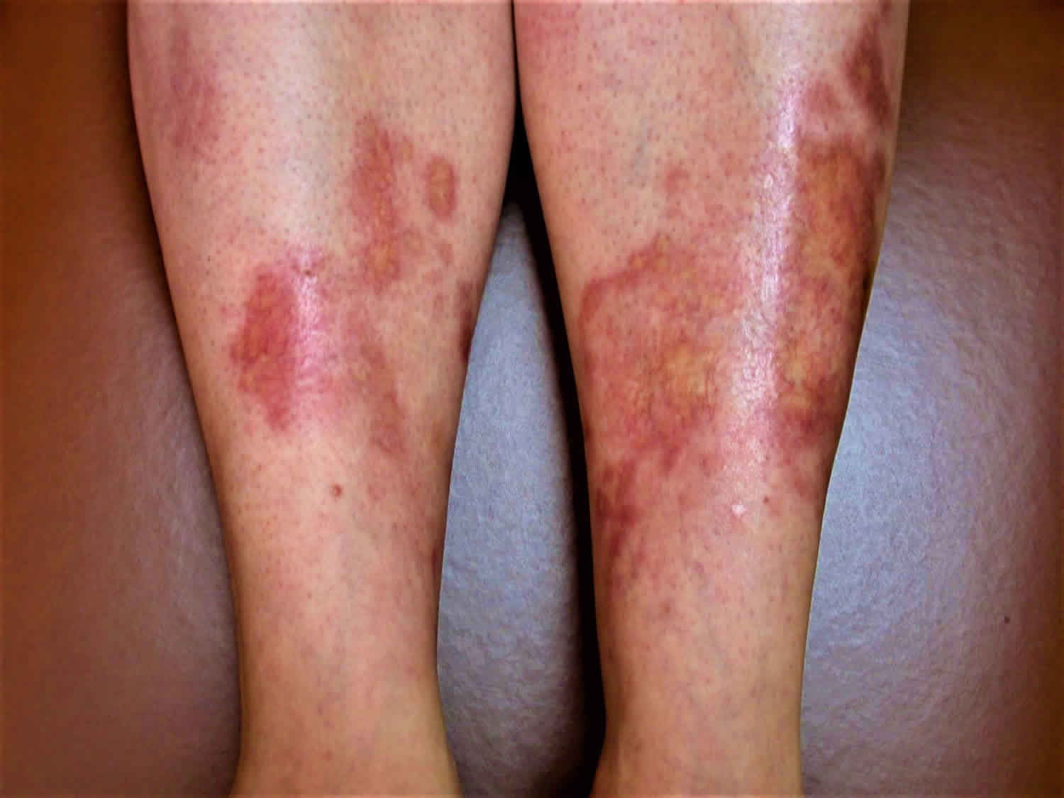
Figure 1. Necrobiosis lipoidica diabeticorum leg
Necrobiosis lipoidica diabeticorum causes
The cause of necrobiosis lipoidica remains unknown. Elements of a vascular disturbance involving immune complex deposition or microangiopathic changes leading to collagen degeneration have been the most common theories 4. Some investigators consider necrobiosis lipoidica to be primarily a disease of collagen, with inflammation occurring as a secondary event.
Necrobiosis lipoidica is thought to be linked to blood vessel inflammation related to autoimmune factors. This damages proteins in the skin (collagen).
People with type 1 diabetes are more likely to get necrobiosis lipoidica diabeticorum than those with type 2 diabetes. Women are more affected than men. Smoking increases the risk for necrobiosis lipoidica diabeticorum. Less than one half of one percent of those with diabetes suffer from this problem.
O Cohen et al reported that necrobiosis lipoidica diabeticorum is usually associated with poor glycemic control, and that improved management of blood glucose levels could improve or prevent the disorder.
Research has also been conducted investigating the effects of smoking in relation to necrobiosis lipoidica diabeticorum, with WF Kelly et al reporting that smoking was prevalent in diabetic patients with necrobiosis lipoidica diabeticorum. This was also the case for retinopathy and nephropathy.
Other possible theories for causes of necrobiosis lipoidica diabeticorum include trauma and inflammatory and metabolic changes.
Necrobiosis lipoidica diabeticorum symptoms
The legs are the most commonly affected site for necrobiosis lipoidica diabeticorum, but other body parts such as the face, abdomen, and scalp have been reported as affected areas. Initially with necrobiosis lipoidica diabeticorum, red-brown patches or bumps (papules) between 1-3 mm develop over months or years on the shins and lower part of the legs, but these can increase in size and become progressively yellow and shiny. They usually appear in the same areas on opposite sides of the body. They are painless in the early stage.
As the papules become bigger, they flatten down. They develop a shiny yellow brown center with raised red to purplish edges. Veins are visible below the yellow part of the lesions. The lesions are irregularly round or oval with well-defined borders. They can spread and join together to give the appearance of a patch.
Lesions can also occur on the forearms. Rarely, they may occur on the stomach, face, scalp, palms, and soles of the feet.
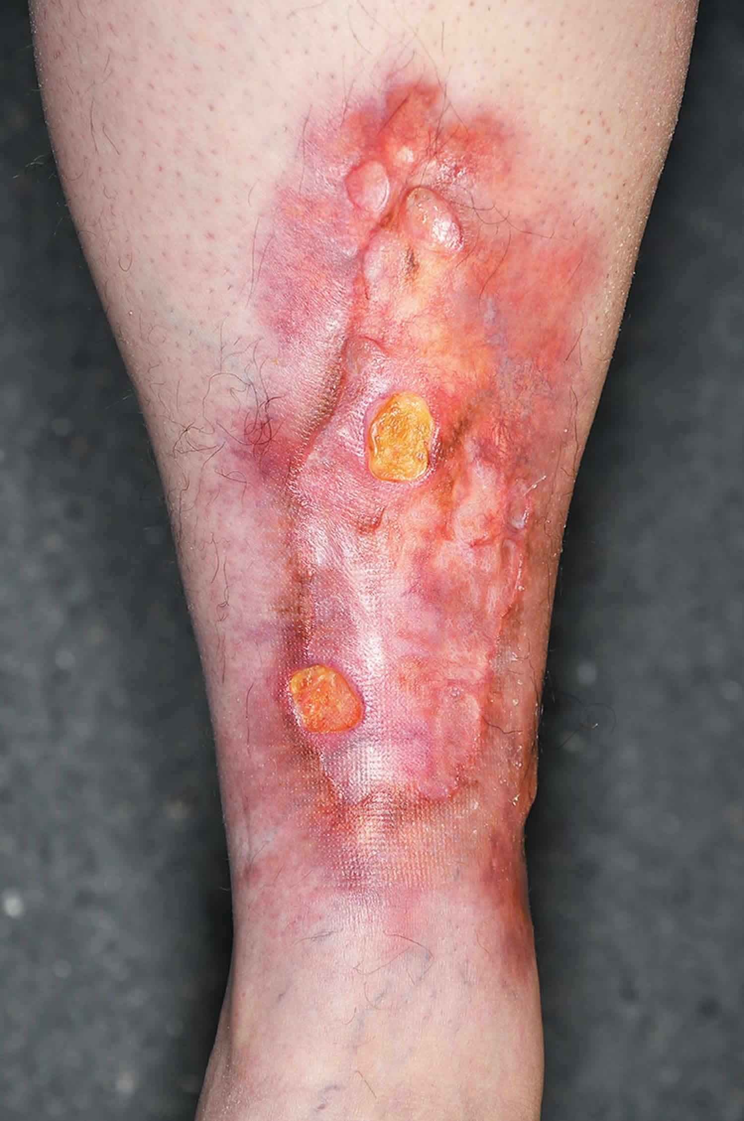
Ulcerations can occur following trauma, which may be associated with some pain. Nodules also may develop. The area may become very itchy and painful.
The Koebner phenomenon – in which new psoriasis plaques form following a skin infection or injury – has been established in patients with necrobiosis lipoidica diabeticorum.
Necrobiosis lipoidica diabeticorum is different from ulcers that can occur on the feet or ankles in people with diabetes.
When legions are multiple and bilateral, they may either be painless due to cutaneous nerve damage, which is often the case, or they can be extremely painful.
Necrobiosis lipoidica diabeticorum diagnosis
Although the diagnosis often is based on clinical examination, a biopsy should be performed to differentiate necrobiosis lipoidica from conditions with similar clinical appearances, including granuloma annulare and necrobiotic xanthogranuloma. A skin biopsy is used to diagnose necrobiosis lipoidica diabeticorum, which can also be the first sign of diabetes in some patients. This is why a patient’s glucose tolerance may also be checked 5.
If there is a concern for venous disease or peripheral arterial disease, further studies should be considered. Baseline blood work should include a fasting blood glucose or glycosylated hemoglobin to screen for diabetes or assess glycemic control in patients known to have diabetes. If these are not diagnostic, they should be repeated on a yearly basis, as necrobiosis lipoidica can be the first presentation of diabetes.
When legions appear on the face, scalp or upper arms, necrobiosis lipoidica diabeticorum is less likely to be correctly diagnosed.
The differential diagnosis includes mainly granuloma annulare, necrobiotic xanthogranuloma, sarcoidosis, diabetic dermopathy, and lipodermatosclerosis. Lesions of granuloma annulare and sarcoidosis generally do not exhibit the same degree of atrophy, telangiectasias or yellow-brown color. Additionally, the appearance of lipid and decreased amount of mucin in necrobiosis lipoidica helps to differentiate from granuloma annulare 6.
Necrobiosis lipoidica diabeticorum treatment
No treatment has proven to be effective. In patients with diabetes mellitus, control of blood glucose usually does not have a significant effect on the course and does not improve symptoms. In the absence of ulceration or symptoms, it is reasonable not to treat necrobiosis lipoidica given that up to 17% of lesions may resolve spontaneously. Compression therapy controls edema and promotes healing in patients with associated venous disease or lymphedema.
Treatment may include:
- Corticosteroid creams
- Injected corticosteroids
- Drugs that suppress the immune system
- Anti-inflammatory drugs
- Medicines that improve blood flow
- Hyperbaric oxygen therapy may be used to increase the amount of oxygen in the blood to promote healing of ulcers
- Phototherapy, a medical procedure in which the skin is carefully exposed to ultraviolet light
- Laser therapy
When ulcerations are present, proper wound care principles are important. First-line therapy includes potent topical corticosteroids for early lesions and intralesional corticosteroids injected into the active borders of established lesions. For inactive, atrophic lesions, topical steroids should be avoided as they may exacerbate the atrophy and increased risk of new ulcerations.
Ultraviolet (UV) light therapy is used for various inflammatory dermatoses. PUVA decreases the actively inflamed borders but has no clinical effects on atrophic scars.
Calcineurin inhibitors inhibit T-cell activation by blocking calcineurin resulting in both anti-inflammatory and immunomodulatory effects. Topical tacrolimus has been shown to be effective in resolving ulceration associated with necrobiosis lipoidica.
TNF is an important regulator of the formation of granulomas. The monoclonal antibodies adalimumab and infliximab bind directly to soluble TNF-alpha to prevent its action. Etanercept is a fusion protein made up of the Fc portion of human IgG1 and TNF receptors that also inhibit TNF function. Both etanercept and infliximab have been repeatedly effective as monotherapy for ulcerating necrobiosis lipoidica.
In severe cases, the lesion may be removed by surgery, followed by moving (grafting) skin from other parts of body to the operated area.
Necrobiosis lipoidica diabeticorum home remedy
During treatment, monitor your glucose level as instructed. Avoid injury to the area to prevent the lesions from turning into ulcers.
If you develop ulcers, follow steps on how to take care of the ulcers.
If you smoke, you will be advised to quit. Smoking can slow healing of the lesions.
Necrobiosis lipoidica diabeticorum prognosis
Necrobiosis lipoidica diabeticorum is a long-term disease. Lesions do not heal well and can recur. Ulcers are difficult to treat. The appearance of the skin may take a long time to become normal, even after treatment.
Necrobiosis lipoidica complications
The main and most serious complication is ulceration, which occurs on the legs in about one third of necrobiosis lipoidica cases. In addition, a few cases of squamous cell carcinoma on chronic ulcerative lesions of necrobiosis lipoidica have been described 7.
Necrobiosis lipoidica diabeticorum can rarely result in skin cancer (squamous cell carcinoma).
Those with necrobiosis lipoidica diabeticorum are at increased risk for:
- Diabetic retinopathy
- Diabetic nephropathy
- Lepe K, Salazar FJ. Necrobiosis Lipoidica. [Updated 2019 Mar 14]. In: StatPearls [Internet]. Treasure Island (FL): StatPearls Publishing; 2019 Jan-. Available from: https://www.ncbi.nlm.nih.gov/books/NBK459318
- Erfurt-Berge C, Seitz AT, Rehse C, Wollina U, Schwede K, Renner R. Update on clinical and laboratory features in necrobiosis lipoidica: a retrospective multicentre study of 52 patients. Eur J Dermatol. 2012 Nov-Dec;22(6):770-5
- Pavlovic MD, Milankovic T, Dinic M, et al. The prevalence of cutaneous manifestations in young patients with type 1 diabetes mellitus. Diabetes Care 2007;30:1964–67
- Sibbald C, Reid S, Alavi A. Necrobiosis Lipoidica. Dermatol Clin. 2015 Jul;33(3):343-60.
- Muller SA, Winkelmann RK. Necrobiosis lipoidica diabeticorum: a clinical and pathological investigation of 171 cases. Arch Dermatol 1966;93:272–81
- Hawryluk EB, Izikson L, English JC. Non-infectious granulomatous diseases of the skin and their associated systemic diseases: an evidence-based update to important clinical questions. Am J Clin Dermatol. 2010;11(3):171-81
- Lim C, Tschuchnigg M, Lim J. Squamous cell carcinoma arising in an area of long-standing necrobiosis lipoidica. J Cutan Pathol 2006;33:581–83


