
What is night blindness
Night blindness also called nyctalopia, is poor vision at night or in dim light. Night blindness is not a disease in itself, but instead night blindness is a symptom of some other type of vision problem. Night blindness may cause problems with driving at night. People with night blindness (nyctalopia) often have trouble seeing stars on a clear night or walking through a dark room, such as a movie theater. Night blindness is often worse just after a person is in a brightly lit environment. Milder cases may just have a harder time adapting to darkness.
In some cases, being very nearsighted (myopia) can make it hard to see at night or in low light.
Take safety measures to prevent accidents in areas of low light. Avoid driving a car at night, unless you get your eye doctor’s approval.
Certain cells in the eye’s retina are responsible for allowing you to see in dim light. If these cells are affected by a disease or condition, night blindness occurs.
Some of the eye conditions that can cause night blindness include 1:
- Nearsightedness (myopia) (seeing well up close but not far away)
- Glaucoma (a disease of the optic nerve connecting the eye to the brain)
- Medicine for glaucoma that constricts (narrows) the pupil
- Cataracts (cloudiness of the eye’s naturally clear lens)
- Diabetes (uncontrolled blood sugar levels)
- Retinitis pigmentosa (an eye disease that causes blindness)
- Too little Vitamin A
- Keratoconus (having a cornea that is very steeply curved)
If you aren’t sure whether you have night blindness, consider the following questions:
- Do you find yourself having trouble moving around your house at night, even with small night lights?
- Is driving at night becoming more difficult?
- Do you avoid going outside at night for fear of tripping?
- Do you have trouble recognizing people’s faces in darkened settings?
- Does it take your eyes a long time to adjust to light when coming in from the darkness?
- Similarly, does it take you a long time to adjust to seeing anything in a darkened room?
If you have any concerns about your ability to see in the dark or in dim light, speak with your eye doctor right away. Having a complete eye exam will help identify any condition affecting your vision.
It is important to have a complete eye exam to determine the cause of night blindness, which may be treatable. Call your eye doctor if symptoms of night blindness persist or significantly affect your life.
Treating night blindness depends solely on its cause. If your refractive error is significant, getting a new prescription for your eyeglasses may be all you need for better vision in low light. In some cases, having cataracts removed can be illuminating as far as your vision is concerned. Your ophthalmologist can explain what is causing your night blindness and suggest how to brighten your outlook.
Vitamin A supplements may be helpful if you have a vitamin A deficiency. Ask your eye doctor about it.
Night blindness symptoms
Night blindness doesn’t mean you are completely unable to see at night, but that your vision is poorer at night or in poor light. Night blindness is not a disease in itself, but instead is a symptom of some other type of vision problem (see causes of night blindness below).
What causes night blindness
The causes of night blindness fall into 2 categories: treatable and nontreatable.
Treatable causes:
- Cataracts (cloudiness of the eye’s naturally clear lens)
- Nearsightedness (myopia)
- Glaucoma (a disease of the optic nerve connecting the eye to the brain)
- Medicine for glaucoma that constricts (narrows) the pupil
- Diabetes (uncontrolled blood sugar levels)
- Keratoconus (having a cornea that is very steeply curved)
- Use of certain drugs e.g., Diethylcarbamazine, Acitretin, Brimonidine
- Vitamin A deficiency (rare)
Nontreatable causes:
- Birth defects e.g., Congenital Stationary Night Blindness
- Retinitis pigmentosa (an eye disease that causes blindness)
To determine what is causing night blindness, an eye doctor will perform a thorough eye exam and may order any of a number of specialized exams.
Glaucoma
Glaucoma is a group of diseases that can damage the eye’s optic nerve and result in vision loss and blindness. Glaucoma is one of the main causes of blindness in the United States. Glaucoma is a leading cause of blindness for people over 60 years old. But blindness from glaucoma can often be prevented with early treatment. Open-angle glaucoma, is the most common form of the disease.
Glaucoma usually happens when the fluid pressure inside the eyes slowly rises, damaging the optic nerve. Often there are no symptoms at first. Without treatment, people with glaucoma will slowly lose their peripheral, or side vision. They seem to be looking through a tunnel. Over time, straight-ahead vision may decrease until no vision remains.
There is no cure, but glaucoma can usually be controlled. Early treatment can help protect your eyes against vision loss. Treatments usually include prescription eyedrops and/or surgery.
Who is at risk for glaucoma?
Some people have a higher than normal risk of getting glaucoma. This includes people who:
- are over age 40
- have family members with glaucoma
- are of African, Hispanic, or Asian heritage
- have high eye pressure
- are farsighted (hyperopia) or nearsighted (myopia)
- have had an eye injury
- use long-term steroid medications
- have corneas that are thin in the center
- have thinning of the optic nerve
- have diabetes, migraines, high blood pressure, poor blood circulation or other health problems affecting the whole body
Talk with an ophthalmologist about your risk for getting glaucoma. People with more than one of these risk factors have an even higher risk of glaucoma.
A comprehensive eye exam can tell if you have glaucoma. People at risk should get eye exams at least every two years. They include:
- African Americans over age 40
- People over age 60, especially Mexican Americans
- People with a family history of glaucoma
In glaucoma, with early treatment, you can often protect your eyes against serious vision loss. Therefore it is very important that it be diagnosed.
Types of glaucoma
There are 2 major types of glaucoma.
Primary open-angle glaucoma
This is the most common type of glaucoma. It happens gradually, where the eye does not drain fluid as well as it should (like a clogged drain). As a result, eye pressure builds and starts to damage the optic nerve. This type of glaucoma is painless and causes no vision changes at first.
Some people can have optic nerves that are sensitive to normal eye pressure. This means their risk of getting glaucoma is higher than normal. Regular eye exams are important to find early signs of damage to their optic nerve.
How does the optic nerve get damaged by open-angle glaucoma?
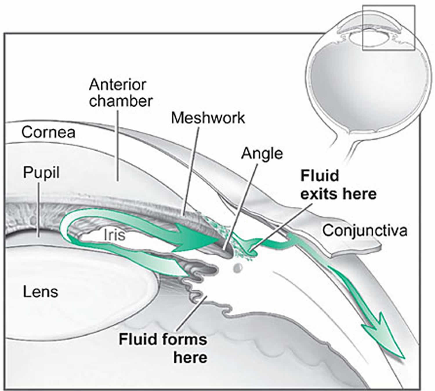
Several large studies have shown that eye pressure is a major risk factor for optic nerve damage. In the front of the eye is a space called the anterior chamber. A clear fluid flows continuously in and out of the chamber and nourishes nearby tissues. The fluid leaves the chamber at the open angle where the cornea and iris meet (see Figure 1 below). When the fluid reaches the angle, it flows through a spongy meshwork, like a drain, and leaves the eye.
In open-angle glaucoma, even though the drainage angle is “open”, the fluid passes too slowly through the meshwork drain. Since the fluid builds up, the pressure inside the eye rises to a level that may damage the optic nerve. When the optic nerve is damaged from increased pressure, open-angle glaucoma-and vision loss—may result. That’s why controlling pressure inside the eye is important.
Another risk factor for optic nerve damage relates to blood pressure. Thus, it is important to also make sure that your blood pressure is at a proper level for your body by working with your medical doctor.
Closed-angle glaucoma
Angle-closure glaucoma (also called “closed-angle glaucoma” or “narrow-angle glaucoma”)
This type happens when someone’s iris is very close to the drainage angle in their eye. The iris can end up blocking the drainage angle. You can think of it like a piece of paper sliding over a sink drain. When the drainage angle gets completely blocked, eye pressure rises very quickly. This is called an acute attack. It is a true eye emergency, and you should call your ophthalmologist right away or you might go blind.
Here are the signs of an acute angle-closure glaucoma attack:
- Your vision is suddenly blurry
- You have severe eye pain
- You have a headache
- You feel sick to your stomach (nausea)
- You throw up (vomit)
- You see rainbow-colored rings or halos around lights
Many people with angle-closure glaucoma develop it slowly. This is called chronic angle-closure glaucoma. There are no symptoms at first, so they don’t know they have it until the damage is severe or they have an attack.
Angle-closure glaucoma can cause blindness if not treated right away.
Figure 1. Glaucoma – fluid pathway is shown in green
Glaucoma signs and symptoms
Open-angle glaucoma
With open-angle glaucoma, there are no warning signs or obvious symptoms in the early stages. As the disease progresses, blind spots develop in your peripheral (side) vision.
Most people with open-angle glaucoma do not notice any change in their vision until the damage is quite severe. This is why glaucoma is called the “silent thief of sight.” Having regular eye exams can help your ophthalmologist find this disease before you lose vision. Your ophthalmologist can tell you how often you should be examined.
Angle-closure glaucoma
People at risk for angle-closure glaucoma usually show no symptoms before an attack. Some early symptoms of an attack may include blurred vision, halos, mild headaches or eye pain. People with these symptoms should be checked by their ophthalmologist as soon as possible. An attack of angle-closure glaucoma includes the following:
- severe pain in the eye or forehead
- redness of the eye
- decreased vision or blurred vision
- seeing rainbows or halos
- headache
- nausea
- vomiting
Normal tension glaucoma
People with “normal tension glaucoma” have eye pressure that is within normal ranges, but show signs of glaucoma, such as blind spots in their field of vision and optic nerve damage.
Glaucoma suspects
Some people have no signs of damage but have higher than normal eye pressure (called ocular hypertension). These patients are considered “glaucoma suspects” and have a higher risk of eventually developing glaucoma. They should be carefully monitored by an ophthalmologist.
Causes of glaucoma
Your eye constantly makes aqueous humor. As new aqueous flows into your eye, the same amount should drain out. The fluid drains out through an area called the drainage angle. This process keeps pressure in the eye (called intraocular pressure or IOP) stable. But if the drainage angle is not working properly, fluid builds up. Pressure inside the eye rises, damaging the optic nerve.
The optic nerve is made of more than a million tiny nerve fibers. It is like an electric cable made up of many small wires. As these nerve fibers die, you will develop blind spots in your vision. You may not notice these blind spots until most of your optic nerve fibers have died. If all of the fibers die, you will become blind.
Glaucoma diagnosis
The only sure way to diagnose glaucoma is with a complete eye exam. A glaucoma screening that only checks eye pressure is not enough to find glaucoma.
During a glaucoma exam, your ophthalmologist (eye specialist) will:
- measure your eye pressure
- inspect your eye’s drainage angle
- examine your optic nerve for damage
- test your peripheral (side) vision
- take a picture or computer measurement of your optic nerve
- measure the thickness of your cornea
Glaucoma treatment
Glaucoma damage is permanent—it cannot be reversed. But medicine and surgery help to stop further damage.
Treating glaucoma successfully is a team effort between you and your doctor. Your ophthalmologist will prescribe your glaucoma treatment. It is up to you to follow your doctor’s instructions and use your eye drops.
Once you are taking medications for glaucoma, your ophthalmologist will want to see you regularly. You can expect to visit your ophthalmologist about every 3–6 months. However, this can vary depending on your treatment needs.
If you have any questions about your eyes or your treatment, talk to your ophthalmologist.
To treat glaucoma, your ophthalmologist may use one or more of the following treatments.
Medication
Glaucoma is usually controlled with eyedrop medicine. Used every day, these eye drops lower eye pressure. Some do this by reducing the amount of aqueous fluid the eye makes. Others reduce pressure by helping fluid flow better through the drainage angle.
It is extremely important to use your glaucoma eye drops exactly as your ophthalmologist tells you to. That includes taking every dose, every day. If you do not do this, you may lose vision.
Also, remember to tell your other doctors which medicines you take for glaucoma. As with any medication, glaucoma eye drops can cause side effects.
Your ophthalmologist may have you take more than one of the following glaucoma eyedrop medicines.
Alpha agonists for glaucoma
Alpha agonists work by reducing the amount of fluid your eye produces. They also increase the amount of fluid that drains out of the eyes. This helps lower eye pressure.
Possible side effects of alpha agonists include:
- red, stinging or painful eyes after using drops
- blurry vision
- allergy (redness, itching, tearing and swelling of the eye)
- a large (dilated) pupil
- headaches
- dry mouth
- feeling tired, weak or dizzy
- an increase in blood pressure
- a fast or irregular heartbeat
- feeling nervous
Do not drive or operate machinery if your glaucoma eye drops make you feel tired or drowsy!
Blurry vision, stinging, and redness may improve with time. But if the side effects still bother you, call your ophthalmologist. He or she may be able to lower your dose or change your medicine. Most side effects go away when the medicine is stopped. Never suddenly quit taking your medicine unless your doctor tells you to.
Beta-blockers for glaucoma
Beta-blockers work by reducing the amount of fluid your eye produces. This helps lower pressure in your eye.
Possible side effects of beta-blockers include:
- red, stinging or painful eyes after using drops
- blurry vision
- breathing problems in people with asthma, emphysema, or COPD
- a slow or irregular heartbeat
- feeling tired
- depression
- dizziness
- a change in sex drive or sexual function
- getting overly tired during exercise
- in people with diabetes, low blood sugar symptoms becoming difficult to notice
Blurry vision, stinging, and redness may improve with time. But if the side effects still bother you, call your ophthalmologist. He or she may be able to lower your dose or change your medicine. Most side effects go away when the medication is stopped. Never suddenly quit taking your medicine unless your doctor tells you to.
Carbonic anhydrase inhibitors for glaucoma
Carbonic anhydrase inhibitors work by reducing the amount of fluid your eye produces. This helps lower eye pressure.
Your ophthalmologist may have you take this medicine as an eye drop or by mouth as a pill.
Possible side effects of carbonic anhydrase inhibitors include:
- stinging eyes
- red eyes
- blurry vision
- a skin rash (especially in people who are allergic to sulfa drugs)
- changes in how things taste to you (especially with carbonated drinks)
- bad taste or upset stomach (nausea)
- feeling tired
- decreased energy
- increase in urination (with the pills)
- tingling around the mouth and fingertips (with the pills)
Blurry vision, stinging, and redness may improve with time. But if the side effects still bother you, call your ophthalmologist. He or she may be able to lower your dose or change your medicine. Most side effects go away when the medication is stopped. Never suddenly quit taking your medicine unless your doctor tells you to.
Miotics for glaucoma
Miotics make your pupil constrict (get smaller), increasing the amount of fluid that drains out of the eye. This helps lower eye pressure.
Possible side effects of miotics include:
- blurred vision
- nearsightedness (trouble focusing on distant objects)
- dim vision with difficulty seeing in the dark or at night (night blindness)
- headache or brow ache (aching around eye)
While very rare, there is the possibility that your retina could detach. This is when the light-sensitive tissue lining the back of the eye pulls away. You would suddenly notice dark specks or spots (floaters) or flashing lights in your vision. If you have these symptoms, call your ophthalmologist immediately.
Side effects may go away after you take the medicine for a while. But if the side effects still bother you, call your ophthalmologist. He or she may be able to lower your dose or change your medicine. Never suddenly quit taking your medicine unless your doctor tells you to.
Prostaglandin analogs for glaucoma
Prostaglandin analogs work by increasing the drainage of fluid out of your eye. This helps lower eye pressure.
Possible side effects of prostaglandin analogs include:
- red, stinging or painful eyes after using drops
- feeling like something is in your eye
- blurry vision
- a permanent change in your eye color (occurs mostly in hazel eyes)
- an increase in thickness, number and length of eyelashes
- darkening of the eyelid
- upper respiratory tract infections, such as colds and flu
- joint aches
- light sensitivity
- eyes gradually sinking deeper into their sockets, keeping eyelids from working properly
Blurry vision, stinging, and redness may improve with time. But if the side effects still bother you, call your ophthalmologist. He or she may be able to lower your dose or change your medicine. Most side effects go away when the medication is stopped. Never suddenly quit taking your medicine unless your doctor tells you to.
All medications can have side effects. Some drugs can cause problems when taken with other medications. It is important to give your doctor a list of every medicine you take regularly. Be sure to talk with your ophthalmologist if you think you may have side effects from glaucoma medicine.
Never change or stop taking your glaucoma medications without talking to your ophthalmologist. If you are about to run out of your medication, ask your ophthalmologist if you should have your prescription refilled.
Laser eye surgery
There are two main types of laser eye surgery to treat glaucoma. They help aqueous drain from the eye. These procedures are usually done in the ophthalmologist’s office or an outpatient surgery center.
- Trabeculoplasty. This surgery is for people who have open-angle glaucoma. The eye surgeon uses a laser to make the drainage angle work better. That way fluid flows out properly and eye pressure is reduced.
- Iridotomy. This is for people who have angle-closure glaucoma. The ophthalmologist uses a laser to create a tiny hole in the iris. This hole helps fluid flow to the drainage angle.
Operating room eye surgery
Some glaucoma surgery is done in an operating room. It creates a new drainage channel for the aqueous humor to leave the eye.
- Trabeculectomy. This is where your eye surgeon creates a tiny flap in the sclera (white of your eye). He or she will also create a bubble (like a pocket) in the conjunctiva called a filtration bleb. It is usually hidden under the upper eyelid and cannot be seen. Aqueous humor will be able to drain out of the eye through the flap and into the bleb. In the bleb, the fluid is absorbed by tissue around your eye, lowering eye pressure.
- Glaucoma drainage devices. Your ophthalmologist may implant a tiny drainage tube in your eye. It sends the fluid to a collection area (called a reservoir). Your eye surgeon creates this reservoir beneath the conjunctiva (the thin membrane that covers the inside of your eyelids and white part of your eye). The fluid is then absorbed into nearby blood vessels.
Retinitis pigmentosa
Retinitis pigmentosa refers to a group of inherited retinal diseases causing retinal degeneration. The retina is a thin piece of tissue lining the back of the eye. The retina converts light into electrical signals that the brain interprets as vision. People with retinitis pigmentosa experience a gradual decline in their vision, because photoreceptor cells in the retina degenerate.
An estimated 100,000 people in the U.S. have retinitis pigmentosa, mainly caused by gene mutations (variations) inherited from one or both parents.
Retinitis pigmentosa is characterized by diffuse progressive dysfunction of predominantly rod photoreceptors, with subsequent degeneration of cone photoreceptors, and retinal pigment epithelium (RPE) 2. In the early stages of retinitis pigmentosa, rods are more severely affected than cones. As the rods die, people experience night blindness and a progressive loss of the visual field, the area of space that is visible at a given instant without moving the eyes. The loss of rods eventually leads to a breakdown and loss of cones. In the late stages of retinitis pigmentosa, as cones die, people tend to lose more of the visual field, developing ìtunnel vision. They may have difficulty performing essential tasks of daily living such as reading, driving, walking without assistance, or recognizing faces and objects.
Pathologic findings of an enucleated eye in a patient with autosomal recessive retinitis pigmentosa showed that the rod and cone outer segments were shortened and disorganized in the patient’s best field of vision, while in the area of visual loss; there was total loss of outer segments and a decrease in photoreceptors number 3. Two types of pigmented cells were found invading the retina: typical retinal pigment epithelium (RPE) cells that were migrating away from the retinal pigment epithelial layer, and macrophage-like cells that contained melanin. These changes were thought to be a reactive response to photoreceptor damage, since the retinal pigment epithelium (RPE) appeared relatively normal morphologically in areas of early photoreceptor involvement. A recent review described histopathologic findings in 10 patients with autosomal dominant retinitis pigmentosa, including poorly organized, shortened or absent outer segments with shortened inner segments. Inclusion bodies and/or perinuclear cytoplasmic membranous swirls were found in three cases 4. Visual impairment usually manifest as night blindness and progressive visual field loss. Retinitis pigmentosa prevalence is 1:3000 to 1:5000 5. Retinitis pigmentosa may be seen in isolation (typical retinitis pigmentosa) or in association with systemic disease.
Forms of retinitis pigmentosa and related diseases include Usher syndrome, Leber congenital amaurosis, and Bardet-Biedl syndrome, among others.
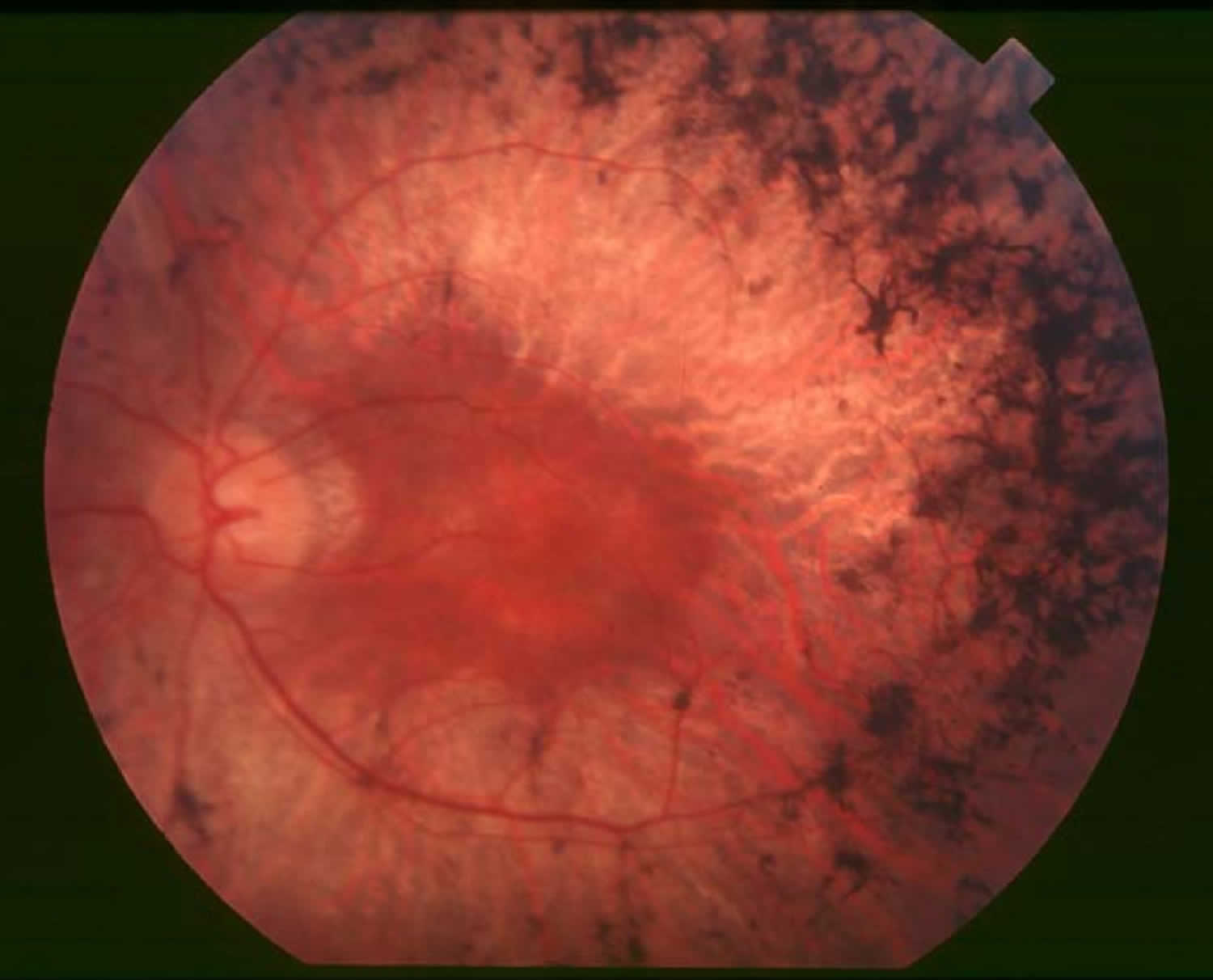
Figure 2. Retinitis Pigmentosa
Retinitis pigmentosa causes
Retinitis pigmentosa is actually a genetically determined degeneration (dystrophy) and not an inflammatory disorder. Since retinitis pigmentosa is a collection of many different genetic disorders, the cause is quite variable. However, the final common pathway appears to be photoreceptor cell death by apoptosis (rods followed by cones).
Mutations in more than 60 genes are known to cause nonsyndromic retinitis pigmentosa 6. More than 20 of these genes are associated with the autosomal dominant form of the disorder. Mutations in the RHO gene are the most common cause of autosomal dominant retinitis pigmentosa, accounting for 20 to 30 percent of all cases. At least 35 genes have been associated with the autosomal recessive form of the disorder. The most common of these is USH2A; mutations in this gene are responsible for 10 to 15 percent of all cases of autosomal recessive retinitis pigmentosa. Changes in at least six genes are thought to cause the X-linked form of the disorder. Together, mutations in the RPGR and RP2 genes account for most cases of X-linked retinitis pigmentosa.
The genes associated with retinitis pigmentosa play essential roles in the structure and function of specialized light receptor cells (photoreceptors) in the retina. The retina contains two types of photoreceptors, rods and cones. Rods are responsible for vision in low light, while cones provide vision in bright light, including color vision.
Mutations in any of the genes responsible for retinitis pigmentosa lead to a gradual loss of rods and cones in the retina. The progressive degeneration of these cells causes the characteristic pattern of vision loss that occurs in people with retinitis pigmentosa. Rods typically break down before cones, which is why night vision impairment is usually the first sign of the disorder. Daytime vision is disrupted later, as both rods and cones are lost.
Some of the genes associated with retinitis pigmentosa are also associated with other eye diseases, including a condition called cone-rod dystrophy. Cone-rod dystrophy has signs and symptoms similar to those of retinitis pigmentosa. However, cone-rod dystrophy is characterized by deterioration of the cones first, followed by the rods, so daylight and color vision are affected before night vision.
Retinitis pigmentosa inheritance pattern
Retinitis pigmentosa often has an autosomal dominant inheritance pattern, which means one copy of an altered gene in each cell is sufficient to cause the disorder. Most people with autosomal dominant retinitis pigmentosa have an affected parent and other family members with the disorder.
Retinitis pigmentosa can also have an autosomal recessive pattern of inheritance, which means both copies of a gene in each cell have mutations. The parents of an individual with an autosomal recessive condition each carry one copy of the mutated gene, but they typically do not show signs and symptoms of the condition.
This condition can also be inherited in an X-linked pattern. The genes associated with X-linked retinitis pigmentosa are located on the X chromosome, which is one of the two sex chromosomes. In males (who have only one X chromosome), one altered copy of the gene in each cell is sufficient to cause the condition. In females, (who have two X chromosomes), mutations usually have to occur in both copes of the gene to cause the disorder. However, at least 20 percent of females who carry only one mutated copy of the gene develop retinal degeneration and associated vision loss. In most cases, males experience more severe symptoms of the disorder than females. A characteristic of X-linked inheritance is that fathers cannot pass X-linked traits to their sons.
In 10 to 40 percent of all cases of retinitis pigmentosa, only one person in a family is affected. In these families, the disorder is described as simplex. It can be difficult to determine the inheritance pattern of simplex cases because affected individuals may have no affected relatives or may be unaware of other family members with the disease. Simplex cases can also result from a new gene mutation that is not present in other family members.
Retinitis pigmentosa symptoms
Symptoms depend on whether rods or cones are initially involved. In most forms of retinitis pigmentosa, rods are affected first. Because rods are concentrated in the outer portions of the retina and are triggered by dim light, their degeneration affects peripheral and night vision. When the disease progresses and cones become affected, visual acuity, color perception, and central vision are diminished.
Night blindness is one of the earliest and most frequent symptoms of retinitis pigmentosa. People with mainly cone degeneration, however, first experience decreased central vision and reduced ability to discriminate colors and perceive details.
Patients typically presents with night vision problems (unable to see in the dark or slow to adjusting to dark), progressive peripheral vision restriction, and tunnel vision at later stage of the disease.
Retinitis pigmentosa is typically diagnosed in adolescents and young adults. It is a progressive disorder. The rate of progression and degree of visual loss varies from person to person. Most people with retinitis pigmentosa are legally blind by age 40, with a central visual field of less than 20 degrees in diameter. It is a genetic disorder and, therefore, is almost always inherited.
It is rare for patients to lose all vision in both eyes. In a large study involving close to 1,000 patients with retinitis pigmentosa and Usher Syndrome at age 45 or older, one fourth of the patients had a visual acuity of 20/200 or worse in both eyes, and more than half had a visual acuity of 20/40 or better in at least one eye. Only 0.5% of patients were completely blind in both eyes 7. In one study, about 50% of retinitis pigmentosa patients reported having headaches, and 35% of retinitis pigmentosa patients reported light flashes 8.
How does retinitis pigmentosa progress?
The symptoms of retinitis pigmentosa typically appear in childhood. Children often have difficulty getting around in the dark. It can also take abnormally long periods of time to adjust to changes in lighting. As their visual field becomes restricted, patients often trip over things and appear clumsy. People with retinitis pigmentosa often find bright lights uncomfortable, a condition known as photophobia. Because there are many gene mutations that cause the disorder, its progression can differ greatly from person to person. Some people retain central vision and a restricted visual field into their 50s, while others experience significant vision loss in early adulthood. Eventually, most individuals with retinitis pigmentosa will lose most of their sight.
How is retinitis pigmentosa diagnosed?
Retinitis pigmentosa is diagnosed in part through an examination of the retina. An eye care professional will use an ophthalmoscope, a tool that allows for a wider, clear view of the retina. This typically reveals abnormal, dark pigment deposits that streak the retina. These pigment deposits are in part why the disorder was named retinitis pigmentosa. Other tests for retinitis pigmentosa include:
Diagnostic procedures
- Full-Field Electroretinogram (ERG): Electroretinogram measures the electrical potential generated by rods and cones after a light stimulus and is essential in the diagnosis of retinitis pigmentosa. The most important parameters being measured include a- and b-wave amplitudes and implicit times. In early stages of the disease, there is reduction in a- and b-wave amplitudes but implicit time can be prolonged or normal. Patients with advanced stages have non-detectable electroretinogram.
- Dark adaptometry (DA): Visual threshold is the minimum intensity of light that will stimulate the rods or cones to elicit a subjective response. Dark adaptometry measures the absolute threshold of rods at given time intervals as the retina adapts to the dark. In retinitis pigmentosa, there is increased absolute rod threshold and dark adaptation is usually prolonged. This test maybe useful in detecting early cases 9.
- Visual field: Kinetic perimetry with Goldmann perimeter characteristically shows a ring scotoma in the mid-periphery of the visual field. They usually start as a group of isolated scotomas around 20 degrees from fixation, and gradually coalesce to form a partial followed by a complete ring. The outer edge of the ring expands relatively quickly to the periphery, while the inner edge constricts slowly toward fixation. Patients often have good central vision from a small central island (“tunnel vision”) until their 50’s or 60’s 10. Visual field testing is useful in monitoring the progression of disease and document the status of legal blindness.
- Electrooculogram (EOG) is a measurement of standing potential between the cornea and the retina and is a measurement of function of the retinitis pigment epitehlium and photoreceptors. It is usually abnormal in retinitis pigmentosa. However, electroretinogram is considered a more sensitive test for detection of photoreceptor function and consequently electrooculogram is not routinely done.
- Optical coherence tomography (OCT): Optical coherence tomography is a quick, inexpensive, and widely available tool to detect cystic macular lesions, epiretinal membrane, and vitreomaular traction syndrome observed in some retinitis pigmentosa patients with decreased central vision. One study also showed mild inner retinal layer thinning and severe outer retinal layer thinning using spectral domain optical coherence tomography 11
- Fluorescein angiography: Fluorescein angiography may have a role in documenting early deterioration of the retinal pigment epithelium and especially in female carriers of X-linked retinitis pigmentosa. It has a role in patients with cystic macular lesions and exudative vasculopathy.
Laboratory test
- Genetic testing can be helpful in confirming the diagnosis. It also helps assess the risk of passing retinitis pigmentosa from parent to offspring.
- Other laboratory testings that can be helpful in differentiating atypical cases of retinitis pigmentosa from other ocular disorders include serologies for syphilis, serum ornithine level or ornithine-lysine ratio (for gyrate atrophy of the retina and choroid), and serum phytanic acid level (for Refsum disease).
Retinitis pigmentosa treatment
Many treatments have been explored without proven benefit for the isolated forms of retinitis pigmentosa 12. These include various vitamins and minerals, vasodilators, tissue therapy with placental extract, cortisone, cervical sympathectomy, injections of a hydrolysate of yeast RNA, ultrasound, transfer factor, dimethyl sulfoxide, ozone, muscle transplants, and subretinal injections of fetal retinal cells. None of the above treatments were conducted in randomized, controlled clinical trials. It is important to note that anecdotal treatment with subjective improvement of visual function should be interpreted with caution due to fluctuation in visual acuity and visual fields in this disease. ERG (electroretinogram) is a better objective measure of remaining retinal function. Any potential therapy will likely require several years of follow-up to assess efficacy due to the nature of slow progression of this disease.
Medical therapy
Controversies exist regarding the use of high dose vitamin A, docosahexaenoic acid (DHA), and lutein to slow the progression of retinitis pigmentosa. Berson et al. 13 conducted three large randomized, controlled, double-masked trials. In the first study, 601 adult patients were randomized to one of four treatment groups: vitamin A, 15,000 IU/day plus vitamin E 3 IU/day; vitamin A 75 IU/day plus vitamin E, 3 IU/day; vitamin A, 15,000 IU/day plus vitamin E, 400 IU/day; and vitamin A, 75 IU/day plus vitamin E, 400 IU/day. The main outcome variable was the 30-Hz cone flicker ERG (electroretinogram). In summary, patients who are on the higher dose of vitamin A had the slowest annual rate of decline in remaining ERG (electroretinogram) amplitude (8.3% of decline per year) while those on high dose vitamin E had the fastest (11.8%). The results were more significant in the cohort with higher amplitudes to start with (i.e., > 0.68 μV).
In the second study, patients who were given vitamin A palmitate 15,000 IU/day were randomized to either docosahexaenoic acid (DHA) capsules (1200 mg/day) or control fatty acid capsules. The main outcome variable was the total point score of the 30-2 Humphrey visual field. Overall, DHA supplementation by capsules did not slow the course of retinitis pigmentosa over a 4-year interval. However, for those who are taking vitamin A for the first time, a subgroup analysis concluded DHA supplement slowed the rate of visual field loss and log ERG amplitude loss in years 1 and 2, but not in years 3 and 4 after the start of treatment.
In the third study, they evaluated the supplemental effects of lutein 12 mg/day combined with high dose vitamin A and high dietary intake of DHA on the rate of retinitis pigmentosa visual field loss. The investigators reported no difference between groups in the rate of decline in the total point score for the HFA 30-2 program (primary outcome measure, p=0.66), nor loss of HFA 30-2 plus 60-4 total point score, logERG amplitude, and logMAR visual acuity (secondary outcomes). However, they did report a significant effect of treatment on the rate of decline for the HFA 60-4 total point score.
Based on these studies, the authors 13 concluded that patients with retinitis pigmentosa would benefit from taking 12 mg of lutein per day in addition to 15,000 IU/d of vitamin A and weekly meals of oily fish, of which DHA is a major component. However, there were some debates regarding these recommendations 14. For example, members of the Data and Safety Monitoring Committee from the first study reported that much of the originally reported significant difference was a consequence of pooling the data and could be attributed to early and consistently large differences between the vitamin E group and all of the other groups 15. In the 2nd and 3rd study, conclusions were drawn based on secondary outcomes and subgroup analyses, rather than primary outcome 14. Therefore, the use of high dose vitamin A and other supplements must be weighed against their potential side effects.
The precise mechanism by which vitamin A supplementation provides its benefit is not known. It has been speculated that vitamin A rescues remaining cones, thereby explaining how one supplement may help a group of patients with different rod-specific gene defects. Vitamin E may lead to an adverse effect on the course of retinitis pigmentosa by inhibiting the absorption or transport of vitamin A. DHA is thought to facilitate the release of vitamin A from its carrier protein (interphotoreceptor retinoid binding protein) in the subretinal space.
Other treatment considerations
Patients who develop cystic macular lesions (about 30%) may benefit from oral acetazolamide 16, topical dorzolamide drops 17, and intravitreal steroids in some cases. Anti-VEGF intravitreal injection has also been shown to be effective in a small case series 18. The long-term efficacy of topical dorzolamide in improving the macular cystic lesions in patients with retinitis pigmentosa and Usher syndrome has been been demonstrated in a retrospective series with a mean follow-up of 39 months 19.
Although light deprivation has not been shown to be of benefit in altering the course of retinal degeneration 20, it is generally advisable for patients to use ultraviolet and short-wavelength (blue) blocking sunglasses for outdoor activities. Audiology consults should be considered for patients with possible or known diagnosis of Usher syndrome. Low vision services are designed to benefit those whose ability to function is compromised by visual impairment. A low vision examination may be useful to help optimize the use of remaining visual function. Genetic counseling can provide patients and families with information on the inheritance and implications of their genetic disorders and can help them make informed medical and personal decisions.
Medical follow up
Annual ocular examinations usually are sufficient to measure visual acuity and Goldmann visual field. If medical treatment is initiated, more frequent visits and laboratory blood work may be indicated. For example, patients with red blood cell docosahexaenoic acid (DHA) level of at least 4% of total red blood cell fatty acids has been reported to have, on average, a slower rate of decline of visual field sensitivity than those with lower levels 21. Vitamin A levels and liver function tests should also be done annually if treatment has been initiated.
Treatment of rare forms of retinitis pigmentosa
In patients with hereditary abetalipoproteinemia (Bassen–Kornzweig syndrome), mutations in the gene encoding a microsomal triglyceride transfer protein lead to depletion of vitamin A in the liver and the retina 22. A low-fat diet and supplementation of fat soluable vitamins A, E, and K are recommended. In patients with Refsum disease, they inherit a defective enzyme that can lead to accumulation of excess phytanic acid 23. Findings other than retinopathy include peripheral neuropathy and ataxia. Treatment consists of restricting food items that contain phytanic acid (including animal fats, milk products, and green leafy vegetables containing phytol) while maintaining body weight.
Surgery
There is now an FDA approved Humanitarian Device, called the ARGUS II implant, which may help patients with end-stage retinitis pigmentosa. It is approved for use in patients with bare light to no light perception. It consists of 3 parts: a video recorder, a transmitter and the implant itself. The implant is an epiretinal electrode chip coated in silicone that stimulates the retina electrically. It is connected to a silicone strip that carries the electrodes from the receiver. This strip encircles the eyeball and is surgically sewn onto the sclera. The wireless receiver receives electrical signals from a video recorder which is mounted to glasses on the patient’s face. The video unit converts the video images into electrical impulses which are transmitted to the receiver. The retinal stimulation results in the patient seeing lines or dots of light that indicate edges or objects in the patient’s field of vision. The patient does not see in color and the resolution does not allow for “seeing faces or small details.” Prior research on the ARGUS II showed that patients are better able to find doors, walk along a path and identify the location and movement of objects with the device turned “on” than without the device 24.
In patients with another form of retinitis pigmentosa, Leber’s variant, gene therapy for retinitis pigmentosa epithelium 65 is being performed. This technique requires the injection of the gene into the eye, specifically into the space under the center of the retina (macular subretinal space). Replacement of the gene in younger patients (versus adults) has allowed patients to gain vision. This treatment is produced by Spark Therapeutics.
If the patient develops a cataract, it is generally advisable to defer surgical removal until the patient can no longer read with the better eye. In one study of 30 patients with retinitis pigmentosa, 83% improved by 2 lines on the Snellen visual acuity chart with cataract surgery 25.
Retinitis pigmentosa treatment complications
In general, toxicity from vitamin A treatment is rare. As a safety measure, patients should have a pretreatment assessment of fasting serum vitamin A levels and liver function and annually thereafter. Because of the potential for birth defects, women who are pregnant or planning to conceive are advised not to take high doses of vitamin A (15,000 IU/day). In older adults, long-term vitamin A supplementation has been associated with a decrease in bone density and up to a 1% increased risk of hip fractures 26. Therefore, postmenopausal women and men over the age of 49 who are taking vitamin A should consult with their primary care physician regarding their bone health. Patients with renal failure or renal transplant should not take vitamin A due to excessive renal re-absorption. Finally, vitamin A should not be given to patients on chronic doxycycline because the combination can lead to increased intracranial pressure.
The 5 year study of the ARGUS II Implant supports the long term safety and benefit of the implant for those blind from retinitis pigmentosa 27. A collaborative has also recently published their recommendations to optimize patient outcomes 28. Most common complications are conjunctival erosion and hypotony. It is rare that the implant would require removal.
Retinitis pigmentosa prognosis
Some studies suggest that the rate of progression, age of onset, and eventual visual loss are related to the mode of inheritance. Autosomal dominant retinitis pigmentosa has the best prognosis, with the majority of patients under 30 years having visual acuity of 20/30 or better. X-linked is the most severe form with appreciable impairment of central visual acuity to 20/200 or less by the fifth decade of life. Autosomal recessive and sporadic cases were intermediate in severity 29. In terms of visual field loss, a study of 104 patients with autosomal dominant retinitis pigmentosa shows 93% of patients under age 20, 89% of those from 20-40, and 60% over the age of 40 had a central visual field radius of 10 degrees or greater with the IV4e test object 30.
Living with vision loss
A number of services and devices are available to help people with vision loss carry out daily activities and maintain their independence. In addition to an eye care professional, it’s important to have help from a team of experts, which may include occupational therapists, orientation and mobility specialists, certified low vision therapists, and others.
Children with retinitis pigmentosa may benefit from low vision aids that maximize existing vision. For example, there are special lenses that magnify central vision to expand visual field and eliminate glare. Computer programs that read text are readily available. Closed circuit televisions with a camera can adjust text to suit oneís vision. Portable lighting devices can adjust a dark or dim environment. Mobility training can teach people to use a cane or a guide dog, and eye scanning techniques can help people to optimize remaining vision. Once a child is diagnosed, he or she will be referred to a low vision specialist for a comprehensive evaluation. Parents may also want to meet with the child’s school administrators and teachers to make sure that necessary accommodations are put in place.
For parents of children with retinitis pigmentosa, one challenge is to determine when a child might need to learn to use a cane or a guide dog. Having regular eye examinations to measure the progress of the disorder will help parents make informed decisions regarding low vision services and rehabilitation.
Vitamin A deficiency
Vitamin A deficiency is the leading cause of childhood blindness in the developing world 31. Vitamin A deficiency is significant, particularly in children and pregnant women. Inadequate vitamin A intake is a major cause of vitamin A deficiency in the developing world, while fat malabsorption after intestinal resection and intestinal bypass, particularly bariatric surgery, has become a rare but emerging cause of vitamin A deficiency in developed nations. In the United States, vitamin A deficiency can occur as a result of malnutrition, malabsorption, or poor vitamin metabolism due to liver disease 32. Malabsorption of vitamin A has been documented to occur after bariatric surgery 33.
Vitamin A is a fat‐soluble vitamin, absorbed through the small intestine, which is required for retinal photoreceptor function, immune function, epithelial proliferation, keratinization and cell and tissue differentiation 34. Two important active metabolites of vitamin A are retinal, the active element of visual pigment, and retinoic acid, an intracellular messenger that modulates cell differentiation 35. Previous studies 36 of vitamin A deficiency in patients with short bowel syndrome reported classic ocular symptoms.
Vitamin A is ingested in the form of retinaldehyde from milk, meat, fish, liver, and eggs. It is also ingested as carotene from green leafy vegetables, yellow fruits, and red palm oil. These compounds are stored in the liver in the form of retinyl pamitate. The aldehyde form of vitamin A, retinal or retinaldehyde, combines with the protein opsin in the rods to create rhodopsin, which is a photosensitive pigment. A similar process takes place in the cones. During phototransduction, some retinal is lost so a constant supply of vitamin A is needed. Vitamin A deficiency can therefore lead to night blindness with associated visual field changes and a depressed electroretinography (ERG) 37. Vitamin A is also necessary for the maintenance of specialized epithelial surfaces. In the conjunctiva, loss of goblet cells and squamous cell metaplasia leads to dryness or xerosis. Bitot’s spots are perilimbal gray plaques of keratinized conjunctival debris overlying an area of xerosis 38. A full-thickness liquefactive necrosis of the cornea (keratomalacia) can also occur 33. Finally, retinopathy in the form of yellow or white punctuate dots can be seen in the retinal periphery 37.
Xerophthalmia is a term used to describe the spectrum of ocular disease that can arise from vitamin A deficiency. Classic symptoms of vitamin A deficiency are ocular manifestations, including reversible night blindness, conjunctival and corneal xerosis (dry eyes), corneal ulceration and melting (kerotomalacia) and retinopathy, which can result in permanent blindness.
Vitamin A is also essential for immune function, and affected children are more susceptible to severe infections, such as measles 38.
More than 95% of the US population has adequate levels of vitamin A, defined as serum retinol ≥20 µg/dL or ≥70 µmol/L 39. Vision loss associated with vitamin A deficiency is relatively common in developing countries and a primary cause of preventable blindness in the world. Globally, 190 million preschool-age children and 19.1 million pregnant women are impacted by low serum retinol, according to estimates by the World Health Organization (WHO) 40. In developed countries, vision loss as a result of vitamin A deficiency generally corresponds with liver failure and poor diet 41. Similar to systemic disease, bariatric procedures have demonstrated a high incidence of elemental and micronutrient deficiencies, including vitamin A 42. An estimated 69% of patients following bariatric surgery demonstrate vitamin A deficiency with approximately 10% manifesting clinical signs 43.
Testing for vitamin A deficiency is available in the form of retinol, retinyl pamitate, and retinol binding protein levels. The World Health Organization (WHO) recommends 200,000 IU of vitamin A for 2 days followed by another dose 2 weeks later for severely malnourished children older than 12 years of age with vitamin A deficiency 44; however, more recent case studies have shown that much less supplementation can lead to reversal of symptoms 45. Vitamin A can also be given intramuscularly if there is a concern for severe malabsorption or corneal involvement requiring a faster recovery.
How much vitamin A do you need ?
According to an analysis of data from the 2007–2008 National Health and Nutrition Examination Survey (NHANES), the average daily dietary vitamin A intake in Americans aged 2 years and older is 607 mcg Retinol Activity Equivalents (RAE) 46. Adult men have slightly higher intakes (649 mcg RAE) than adult women (580 mcg RAE). Although these intakes are lower than the RDAs for individual men and women, these intake levels are considered to be adequate for population groups.
The adequacy of vitamin A intake decreases with age in children 47. Furthermore, girls and African-American children have a higher risk of consuming less than two-thirds of the vitamin A RDA than other children 47.
There are a variety of foods rich in vitamin A and provitamin A carotenoids that are available to Americans. Thus, current dietary patterns appear to provide sufficient vitamin A to prevent deficiency symptoms such as night blindness 48.
- The Estimated Average Requirement (EAR) is based on the assurance of adequate stores of vitamin A.
- The Recommended Dietary Allowance (RDA) (average daily level of intake sufficient to meet the nutrient requirements of nearly all (97%–98%) healthy individuals) for men is 900 μg and women is 700 μg Retinol Activity Equivalents (RAE)/day.
- The Tolerable Upper Intake Level (UL) (maximum daily intake unlikely to cause adverse health effects) for adults is set at 3,000 μg/day of preformed vitamin A.
The Recommended Dietary Allowance (RDA) for vitamin A are given as mcg of retinol activity equivalents (RAE) to account for the different bioactivities of retinol and provitamin A carotenoids (see Table 1). Because the body converts all dietary sources of vitamin A into retinol, 1 mcg of physiologically available retinol is equivalent to the following amounts from dietary sources: 1 mcg of retinol, 12 mcg of beta-carotene, and 24 mcg of alpha-carotene or beta-cryptoxanthin. From dietary supplements, the body converts 2 mcg of beta-carotene to 1 mcg of retinol.
Currently, vitamin A is listed on food and supplement labels in international units (IUs) even though nutrition scientists rarely use this measure. Conversion rates between mcg RAE and IU are as follows 49:
- 1 IU retinol = 0.3 mcg RAE
- 1 IU beta-carotene from dietary supplements = 0.15 mcg RAE
- 1 IU beta-carotene from food = 0.05 mcg RAE
- 1 IU alpha-carotene or beta-cryptoxanthin = 0.025 mcg RAE
An RAE cannot be directly converted into an IU without knowing the source(s) of vitamin A. For example, the RDA of 900 mcg RAE for adolescent and adult men is equivalent to 3,000 IU if the food or supplement source is preformed vitamin A (retinol). However, this RDA is also equivalent to 6,000 IU of beta-carotene from supplements, 18,000 IU of beta-carotene from food, or 36,000 IU of alpha-carotene or beta-cryptoxanthin from food. So a mixed diet containing 900 mcg RAE provides between 3,000 and 36,000 IU of vitamin A, depending on the foods consumed.
The amount of vitamin A you need depends on your age and reproductive status. Recommended intakes for vitamin A for people aged 14 years and older range between 700 and 900 micrograms (mcg) of retinol activity equivalents (RAE) per day. Recommended intakes for women who are nursing range between 1,200 and 1,300 RAE. Lower values are recommended for infants and children younger than 14.
However, the vitamin A content of foods and dietary supplements is given on product labels in international units (IU), not mcg RAE. Converting between IU and mcg RAE is not easy. A varied diet with 900 mcg RAE of vitamin A, for example, provides between 3,000 and 36,000 IU of vitamin A depending on the foods consumed. See our Health Professional Fact Sheet on Vitamin A for more details.
For adults and children aged 4 years and older, the U.S. Food and Drug Administration has established a vitamin A Daily Value (DV) of 5,000 IU from a varied diet of both plant and animal foods. DVs are not recommended intakes; they don’t vary by age and sex, for example. But trying to reach 100% of the DV each day, on average, is useful to help you get enough vitamin A.
The Institute of Medicine developed the Recommended Dietary Allowance (RDA) for vitamin A (retinol). The recommended intakes are listed in International Units (IU) in the table, below:
| Age (yrs) | Children | Men | Women | Pregnancy | Lactation |
|---|---|---|---|---|---|
| [Source: Institute of Medicine, 2001. 50] | |||||
| 1 to 3 | 1,000 | ||||
| 4 to 8 | 1,320 | ||||
| 9 to 13 | 2,000 | ||||
| 14 to 18 | 3,000 | 2,310 | 2,500 | 4,000 | |
| 19+ | 3,000 | 2,310 | 2,565 | 4,300 | |
Vitamin A deficiency signs and symptoms
Vitamin A deficiency signs:
- Dry eyes (xerosis)
- Corneal liquification (keratomalacia)
- Retinal punctate lesions
- Visual field defects
- Depressed electroretinography (ERG)
Figure 3. Vitamin A deficiency dry eyes (xerosis) with Bitot’s spot
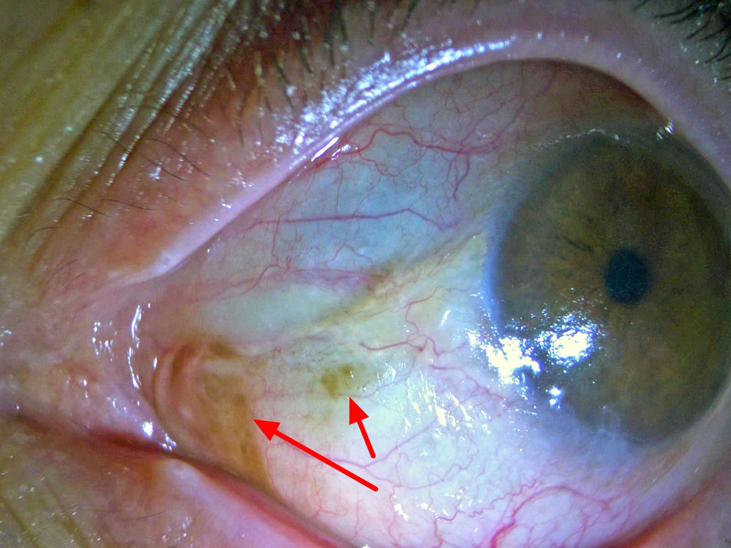
Footnote: Bitot’s spots are considered pathognomic of vitamin A deficiency and correspond to triangular patches of squamous keratinous metaplasia of the conjunctival epithelium, usually near the limbus, typically at the three o’clock and/or nine o’clock positions. The foamy aspect is attributable to colonization by gas-producing bacteria (p.e. Corynebacterium xerosis).
[Source 51]Vitamin A deficiency symptoms:
- Dry eyes
- Night blindness
- Blurry vision
Vitamin A deficiency treatment
- Oral vitamin A 10,000-20,000 IU daily
- Intramuscular vitamin A for severe cases.
How is night blindness treated?
Treatment for night blindness will depend upon its cause. Treatment may be as simple as getting a new eyeglass prescription or switching glaucoma medications, or it may require surgery if the night blindness is caused by cataracts.
If a retinal disease is discovered, the treatment will depend on the type of the disease and will require additional investigation by a retina specialist.
References- Shedding Light on Night Blindness. https://www.aao.org/eye-health/news/shedding-light-on-night-blindness
- Mizuno K, Nishida S: Electron microscopic studies of human retinitis pigmentosa. Part I. Two cases of advanced retinitis pigmentosa. Am J Ophthalmol. 1967;63(4):791-803.
- Bunt-Milam AH, Kalina RE, Pagon RA. Clinical-ultrastructural study of a retinal dystrophy. Invest Ophthalmol Vis Sci. 1983;24(4):458-469.
- Ben-Arie-Weintrob Y, Berson EL, Dryja TP. Histopathologic-genotypic correlations in retinitis pigmentosa and allied diseases. Ophthalmic Genet. 2005;26(2):91-100.
- Weleber RG, Gregory-Evans K. Retinitis pigmentosa and allied disorders. In: Ryan SJ, ed. Retina, 4th edn. Philadelphia, PA: Elsevier; 2006:394-485.
- Retinitis pigmentosa. https://ghr.nlm.nih.gov/condition/retinitis-pigmentosa
- Grover S, Fishman GA, Anderson RJ, et al. Visual acuity impairment in patients with retinitis pigmentosa at age 45 years or older. Ophthalmology. 1999;106(9):1780-1785.
- Heckenlively JR, Yoser SL, Friedman LH et al. Clinical findings and common symptoms in retinitis pigmentosa. Am J Ophthalmol. 1988; 105(5):504-511.
- Alexander KR, Fishman GA. Prolonged rod dark adaptation in retinitis pigmentosa. Br J Ophthalmol. 1984;68(8):561-569.
- Grover S, Fishman GA, Brown J Jr. Patterns of visual field progression in patients with retinitis pigmentosa. Ophthalmology. 1998;105(6):1069-1075.
- Lim JI, Tan O, Fawzi AA, et al: A pilot study of Fourier-domain optical coherence tomography of retinal dystrophy patients. Am J Ophthalmol. 2008;146(3):417-426.
- Berson EL. Retinitis Pigmentosa and Allied Diseases. In: Albert D, Miller J, Azar D, Blodi B, eds. Albert and Jakobiec, 3rd edn. Philadelphia, PA: Elsevier; 2008:Ch. 177.
- Berson EL, Rosner B, Sandberg MA, et al. Clinical trial of lutein in patients with retinitis pigmentosa receiving vitamin A. Arch Ophthalmol. 2010;128(4):403-411.
- Massof RW, Fishman GA. How strong is the evidence that nutritional supplements slow the progression of retinitis pigmentosa? Arch Ophthalmol. 2010;128(4):493-495.
- Norton EWD. A randomized trial of vitamin A and vitamin E supplementation for retinitis pigmentosa [letter to the editor]. Arch Ophthalmol. 1993;111(11):1460.
- Cox SN, Hay E, Bird AC: Treatment of chronic macular edema with acetazolamide. Arch Ophthalmol. 1988; 106(9):1190-1195.
- Grover S, Apushkin MA, Fishman GA: Topical Dorzolamide for the treatment of cystoid macular edema in patients with retinitis pigmentosa. Am J Ophthalmol. 2006; 141(5):850-858.
- Yuzbasioglu E, Artunay O, Rasier R, et al. Intravitreal bevacizumab (Avastin) injection in retinitis pigmentosa. Curr Eye Res. 2009;34(3):231-237.
- Genead MA, Fishman GA. Efficacy of sustained topical dorzolamide therapy for cystic macular lesions in patients with retinitis pigmentosa and Usher syndrome. Arch Ophthalmol. 2010;128(9):1146-1150.
- Berson EL: Light deprivation and retinitis pigmentosa. Vision Res. 1980;20(12):1179-1184.
- Berson EL, Rosner B, Sandberg MA, et al: Further evaluation of docosahexaenoic acid in patients with retinitis pigmentosa receiving vitamin A treatment: Subgroup analyses. Arch Ophthalmol. 2004; 122(9):1306-1314.
- Narcisi TME, Shoulders CC, Chester SA, et al: Mutations of the microsomal triglyceride-transfer-protein gene in abetalipoproteinemia. Am J Hum Genet. 1995;57(6):1298-1310.
- Refsum S: Heredopathia atactica polyneuritiformis: A familial syndrome not hitherto described. Acta Psychiatr Neurol Scand. 1946;38(Suppl):1
- Luo YH, Zhong JJ, da Cruz L. The use of Argus® II retinal prosthesis by blind subjects to achieve localisation and prehension of objects in 3-dimensional space. Graefes Arch Clin Exp Ophthalmol. 2014 Dec 31.
- Stronks HC, Dagnelie G. The functional performance of the Argus II retinal prosthesis. Expert Rev Med Devices. 2014 Jan;11(1):23-30.
- Michaelsson K, Lithell H, Vessby B, Melhus H: Serum retinol levels and the risk of fracture. N Engl J Med. 2003;348(4):287-294.
- deCruz et al. Five-Year Safety and Performance Results from the Argus II Retinal Prosthesis. Ophthalmology. 2016 Oct;123(10):2248-54.
- Ghodasra DH, et al. Worldwide Argus II implantation: recommendations to optimize patient outcomes. BMC Ophthalmol. 2016 May 6;16:52. doi: 10.1186/s12886-016-0225-1.
- Fishman GA, Farber MD, Derlacki DJ. X-linked retinitis pigmentosa. Profile of clinical findings. Arch Ophthalmol. 1988;106(3):369-375.
- Lyness AL, Ernst W, Quinlan MP, et al. A clinical, psychophysical, and electroretinographic survey of patients with autosomal dominant retinitis pigmentosa. Br J Ophthalmol. 1985;69(5):326-339.
- Vitamin A Deficiency and Nyctalopia: 55-year-old male with gradual onset of night blindness. http://webeye.ophth.uiowa.edu/eyeforum/cases/130-vitamin-a-deficiency.htm
- Braunstein A, Trief D, Wang N, Chang S, Tsang S. Vitamin A deficiency in New York. The Lancet 2010;376:267.
- Lee, W. Hamiton S, Harris J, Schwab I. Ocular Complication of Hypovitaminosis A after Bariatric Surgery. Ophthalmology 2005;112:1031-1034.
- Phanachet P, Shantavasinkul PC, Chantrathammachart P, et al. Unusual manifestation of vitamin A deficiency presenting with generalized xerosis without night blindness. Clinical Case Reports. 2018;6(5):878-882. doi:10.1002/ccr3.1475. https://www.ncbi.nlm.nih.gov/pmc/articles/PMC5930185/
- Bates C. J. 1995. Vitamin A. Lancet 345:31–35.
- Cella W., Urbano A. P., Vinhadelli W. S., Donatti M., and Rocha E. M.. 2002. Xerophthalmia secondary to short bowel syndrome. J. Pediatr. Ophthalmol. Strabismus 39:125–127.
- Genead M. Fishman G. Lindeman M. Fundus white spots and acquired night blindness due to vitamin A deficiency. Doc Ophthalmol 2009;119:229–233.
- Smith J, Steinemann TL. Vitamin A deficiency and the eye. Int Ophthalmol Clin 2000;40(4):83-91.
- Second national report on biochemical indicators of diet and nutrition in the U.S. population 2012. https://www.cdc.gov/nutritionreport/pdf/exesummary_web_032612.pdf
- Vitamin A supplements a guide to their use in the treatment and prevention of vitamin A deficiency and xerophthalmia 2nd Edition. http://apps.who.int/iris/bitstream/handle/10665/41947/9241545062.pdf;jsessionid=6212ABDD3AA425BD64F18CAA13C134B9?sequence=1
- Ophthalmic manifestations of vitamin A and D deficiency in two autistic teenagers: case reports and a review of the literature. Duignan E, Kenna P, Watson R, Fitzsimon S, Brosnahan D. Case Rep Ophthalmol. 2015 Jan-Apr; 6(1):24-9. https://www.ncbi.nlm.nih.gov/pmc/articles/PMC4327555/
- Mineral malnutrition following bariatric surgery. Gletsu-Miller N, Wright BN. Adv Nutr. 2013 Sep 1; 4(5):506-17. https://www.ncbi.nlm.nih.gov/pmc/articles/PMC3771134/
- Vitamin a deficiency after gastric bypass surgery: an underreported postoperative complication. Zalesin KC, Miller WM, Franklin B, Mudugal D, Rao Buragadda A, Boura J, Nori-Janosz K, Chengelis DL, Krause KR, McCullough PA. J Obes. 2011; 2011. https://www.ncbi.nlm.nih.gov/pmc/articles/PMC2943134/
- World Health Organization. Management of severe malnutrition: a manual for physicians and other senior health workers. 1999:17-18. http://www.who.int/nutrition/publications/severemalnutrition/en/manage_severe_malnutrition_eng.pdf
- Chae T, Foroozan R. Vitamin A deficiency in patients with a remote history of intestinal surgery. British Journal of Ophthalmology 2006;90:955-956.
- U.S. Department of Agriculture, Agricultural Research Service. What We Eat in America, 2013-2014. https://www.ars.usda.gov/northeast-area/beltsville-md/beltsville-human-nutrition-research-center/food-surveys-research-group/docs/wweia-data-tables/
- Solomons NW. Vitamin A. In: Bowman B, Russell R, eds. Present Knowledge in Nutrition. 9th ed. Washington, DC: International Life Sciences Institute; 2006:157-83.
- Institute of Medicine, US Panel on Micronutrients. Dietary reference intakes for vitamin A, boron, chromium, copper, iodine, iron, manganese, molybdenum, nickel, silicon, vanadium, and zinc. National Academies Press. Washington, DC, 2001. PMID: 25057538 www.ncbi.nlm.nih.gov/pubmed/25057538
- Otten JJ, Hellwig JP, Meyers LD, eds. Dietary Reference Intakes: The Essential Guide to Nutrient Requirements. Washington, DC: The National Academies Press; 2006. https://www.nap.edu/catalog/11537/dietary-reference-intakes-the-essential-guide-to-nutrient-requirements
- Institute of Medicine. Food and Nutrition Board. Dietary Reference Intakes for Vitamin A, Vitamin K, Arsenic, Boron, Chromium, Copper, Iodine, Iron, Manganese, Molybdenum, Nickel, Silicon, Vanadium, and Zinc. Washington, DC: National Academy Press; 2001. https://www.nap.edu/read/10026/chapter/1
- http://ocular-surface.org/photo/bitots-spots-vitamin-deficiency


