What is onychomycosis
Onychomycosis is also known as nail fungus infection, resulting in nail discoloration, thickening, deformity and separation from the nail bed 1. In onychomycosis (fungal nail infection), one, a few, or all nails may be affected. Onychomycosis are common with millions of people suffering from nail fungus infections annually, and the incidence and prevalence of onychomycosis continue to increase worldwide. 2. Onychomycosis are not serious but nail fungus infections can take a long time to treat. Onychomycosis usually affect your toenails but you can get them on your fingernails too. Onychomycosis is increasingly common with increased age, particularly in older individuals (20% of persons older than 60 years and 50% in those older than 70 years) 3. The increased prevalence in older adults is related to peripheral vascular disease, immunologic disorders, and diabetes mellitus. The risk of onychomycosis is 1.9 to 2.8 times higher in persons with diabetes compared with the general population 4. In patients with human immunodeficiency virus infection, the prevalence ranges from 15% to 40% 5. Onychomycosis rarely affects children.
Onychomycosis can be uncomfortable and can lead to cellulitis in older adults 6 and foot ulcers in patients with diabetes 7.
- If you have diabetes you should see a foot specialist because any foot infection can lead to complications.
Onychomycosis affects toenails more often than fingernails because of their slower growth, reduced blood supply, and frequent confinement in dark, moist environments 8. Onychomycosis may occur in patients with distorted nails, a history of nail trauma, genetic predisposition, hyperhidrosis (excessive sweating), concurrent fungal infections, and psoriasis. It is also more common in smokers and in those who use occlusive footwear and shared bathing facilities 3.
Eradication of onychomycosis is key to improving appearance and avoiding these complications, but it is not easily accomplished because nails are made of keratin, which is nonvascular and impermeable to many agents 9. Because of poor drug delivery to nails, results of treatment may not be apparent for a year.
Onychomycosis treatment
Speak to a pharmacist If the look of your nail bothers you or it’s painful.
Your chemist may suggest:
- antifungal nail cream – it can take up to 12 months to cure the infection and doesn’t always work
- nail-softening cream – used for 2 weeks to soften the nail so the infection can be scraped off
The infection is cured when you see healthy nail growing back at the base.
See a doctor if your nail fungus infection:
- is severe and treatment hasn’t worked
- has spread to other nails
Your doctor can prescribe antifungal tablets. You’ll need to take these every day for up to 6 months.
Antifungal tablets can have side effects including:
- headaches
- itching
- loss of taste
- diarrhea
Note: You can’t take antifungal tablets if you’re pregnant or have certain conditions. They can damage your liver.
Badly infected nails sometimes need to be removed. It’s a small procedure done while the area is numbed (under local anaesthetic).
Other treatment
- Laser treatment uses laser to destroy the fungus. It can be expensive. There’s little evidence to show it’s a long-term cure as most studies only follow patients for 3 months.
Onychomycosis signs and symptoms
Onychomycosis may affect one or more toenails and/or fingernails and most often involves the great toenail or the little toenail.
You may have nail fungus if one or more of your nails are:
- Thickened
- Whitish to yellow-brown discoloration
- Brittle, crumbly or ragged
- Distorted in shape
- A dark color, caused by debris building up under your nail
- Smellling slightly foul
In general, toenails are most commonly affected with onychomycosis. If the fingernails are affected, the toenails are usually affected as well. Nails often become thicker and lift from the nail bed (onycholysis) starting at the growing portion of the nail. You might then see debris under the nails and discoloration of the affected area.
- Onychomycosis usually start at the edge of the nail (Figure 1). Onychomycosis often then spread to the middle. The nail becomes discolored and lifts off. The infected nail becomes brittle and pieces can break off. It can cause pain and swelling in the skin around the nail.
- In some forms of onychomycosis, you might see black or white, powdery discoloration on the surface of the nail plate.
- In some forms of onychomycosis, you might see these abnormal changes farther up the finger (near the nailbed), where the nail originates.
Onychomycosis often results from untreated tinea pedis (athlete’s foot) or tinea manuum (tinea or ringworm hand). It may follow an injury to the nail.
Candida infection of the nail plate generally results from paronychia (an infection of the skin around a fingernail or toenail) and starts near the nail fold (the cuticle). The nail fold is swollen and red, lifted off the nail plate. White, yellow, green or black marks appear on the nearby nail and spread. The nail may lift off its bed and is tender if you press on it.
Mold infections are usually indistinguishable from onychomycosis.
Onychomycosis must be distinguished from other nail disorders such as:
- Bacterial infection especially Pseudomonas aeruginosa, which turns the nail black or green.
- Psoriasis.
- Eczema or dermatitis.
- Lichen planus.
- Viral warts.
- Onycholysis (nail lifting of the fingernail or toenail from the nail bed)
- Onychogryphosis (nail thickening and scaling under the nail), common in the elderly.
Clinical features of onychomycosis
White Superficial Onychomycosis
Fungi invade the dorsal nail plate and form colonies that appear as white opaque formations, easily scraped away. The classical form is due to Trichophyton interdigitale, where dermatophytes colonize the most superficial layers of the nail plate without penetrating it (Figure 1), but Fusarium spp. and other molds may cause a white superficial onychomycosis with a deeper nail invasion 10.
Tinea pedis interdigitalis (athlete’s foot) due to Trichophyton interdigitale is common (Figure 2) 11.
Differential diagnosis includes superficial nail fragility due to prolonged wearing of nail polish and transverse toenail leukonychia due to trauma.
Figure 1. White superficial onychomycosis
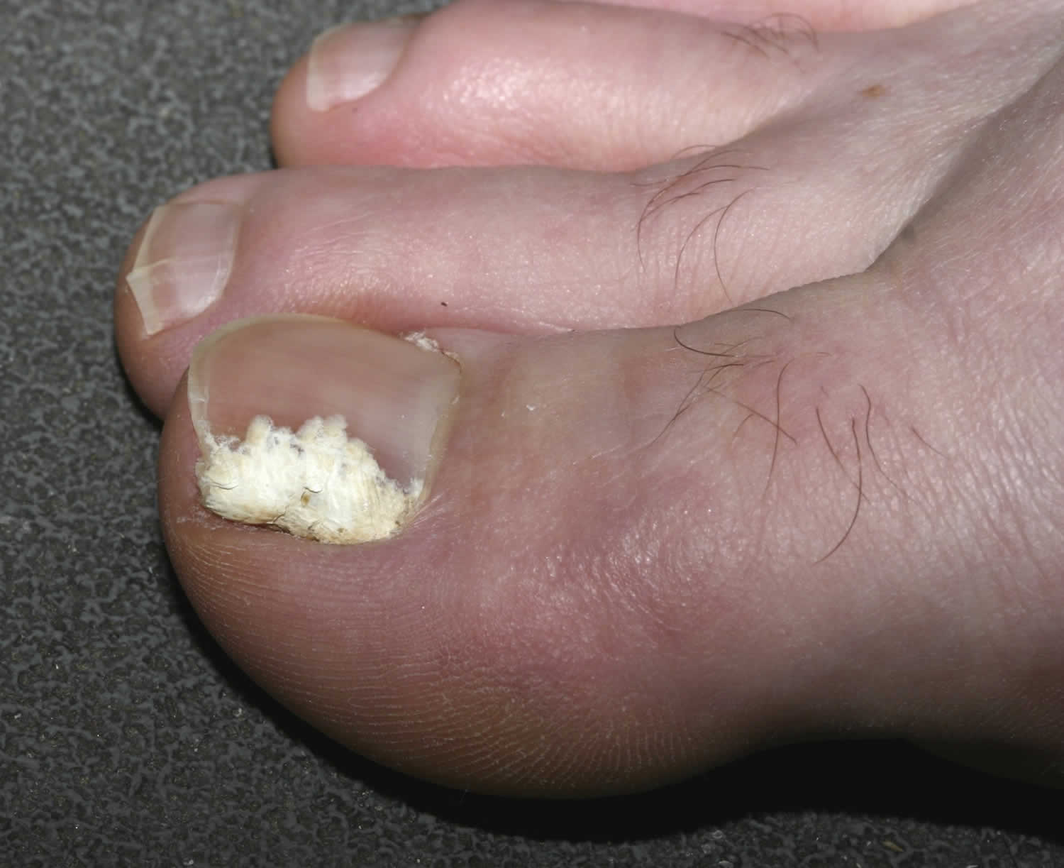
Figure 2. Tinea pedis interdigitalis, often associated with white superficial onychomycosis.
Proximal Subungual Onychomycosis
Fungal elements are typically located in the ventral nail plate, producing a proximal leukonychia. Proximal subungual onychomycosis due to dermatophytes is very rare, and in the past, the form due to Trichophyton rubrum was considered as a sign of HIV infection. It presents as a white area under the proximal nail plate, in the lunula area (Figure 3). Proximal subungual onychomycosis is a common presentation of non-dermatophyte mold infection, especially due to Aspergillus sp. and Fusarium sp., and acute periungual inflammation is often associated. Differential diagnosis includes acute bacterial paronychia and pustular psoriasis of the nail.
Figure 3. Proximal subungual onychomycosis – white discoloration of the proximal nail plate
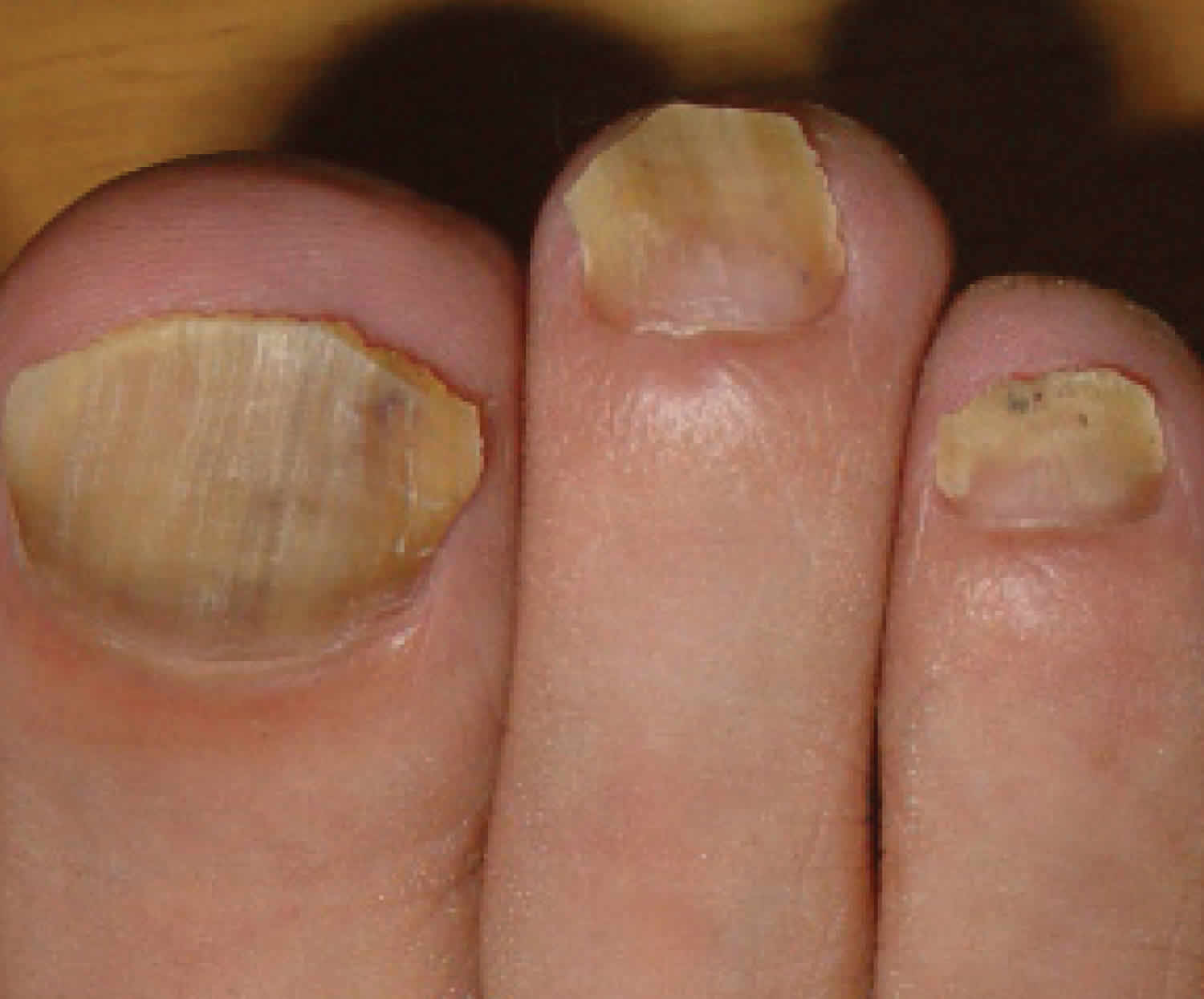
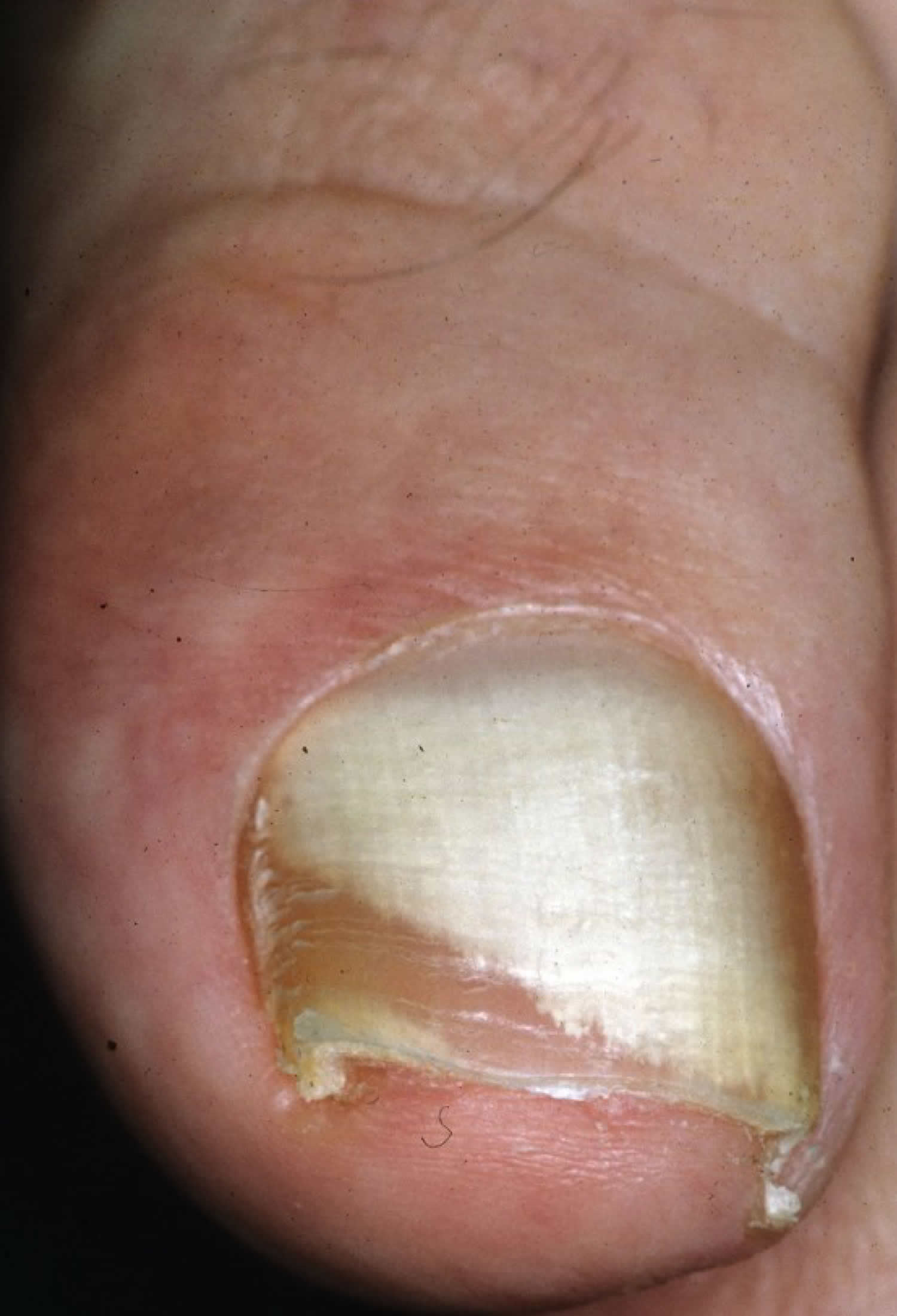
Distal and Lateral Subungual Onychomycosis
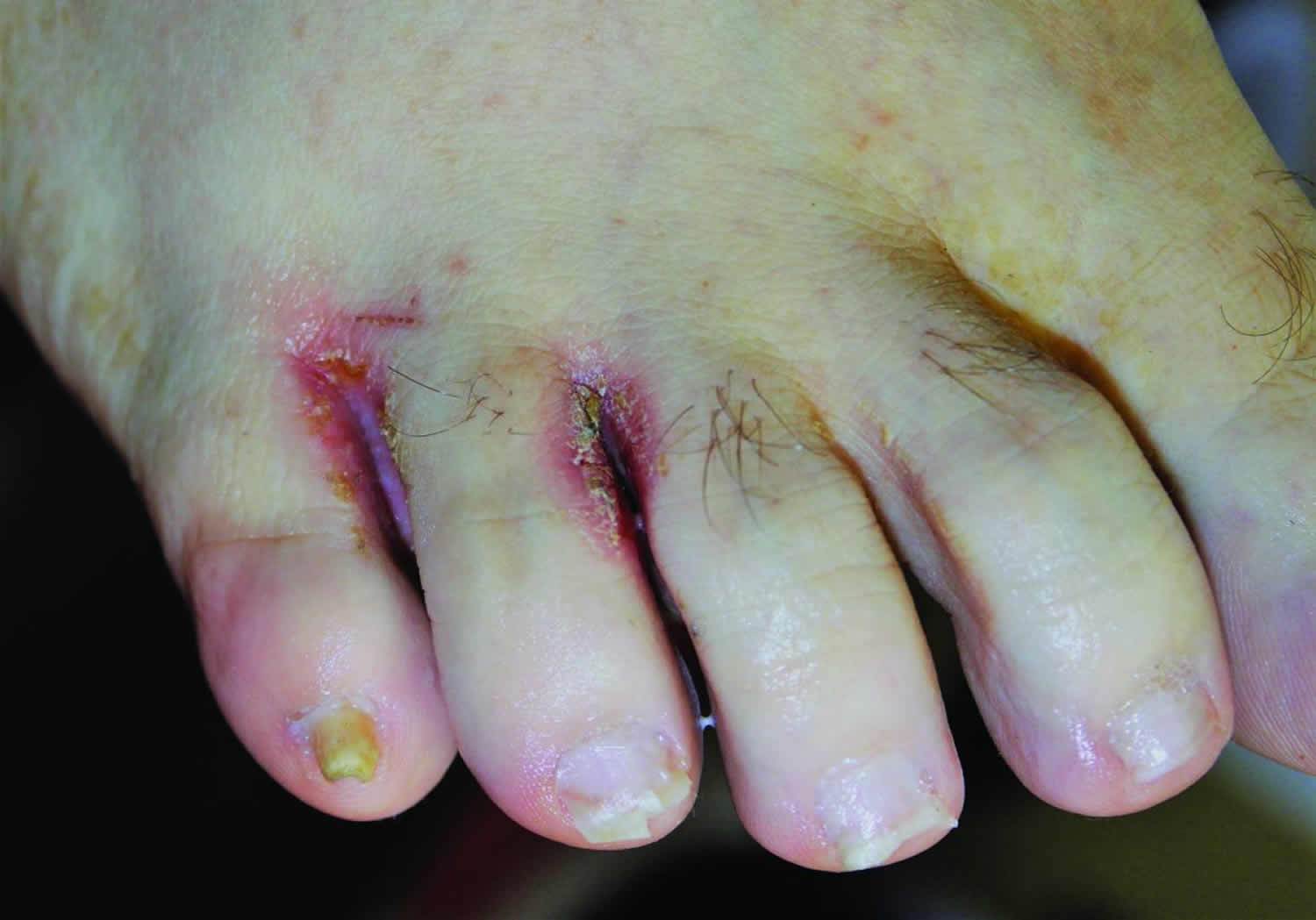
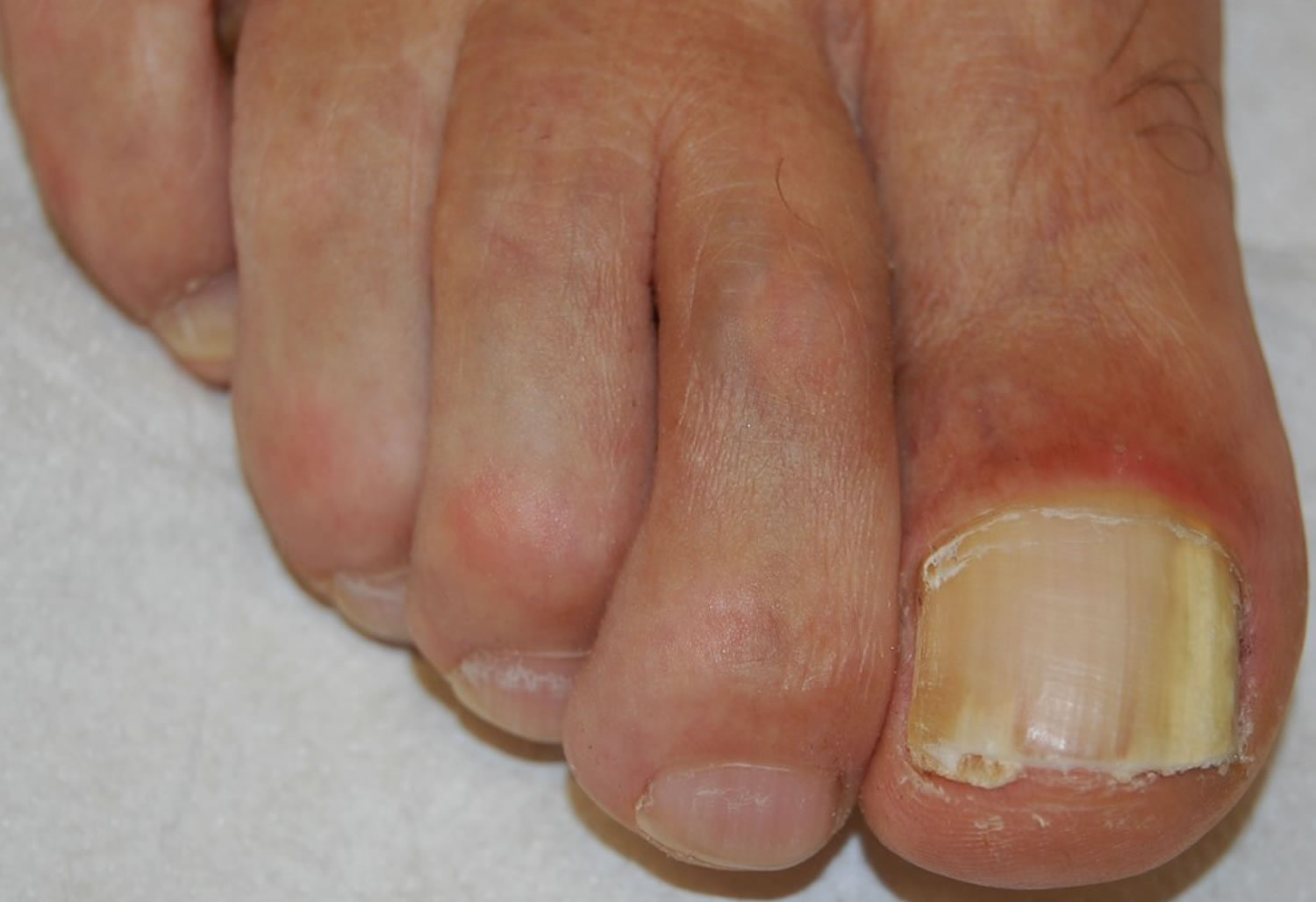
Fungi reach the nail through the hyponychium and invade the undersurface of the nail unit plate spreading proximally. Distal and lateral subungual onychomycosis usually affects one or both of the great toenails and is also usually associated with tinea pedis 11. The nail plate appears yellow-white, is detached due to onycholysis, with distal subungual hyperkeratosis (Figure 4). Less frequently, a brown, black or orange discoloration of the onycholytic nail can be seen (Figure 5). A possible presentation of distal and lateral subungual onychomycosis due to dermatophytes is dermatophytoma, a subungual accumulation of hyphae and scales, scarcely reached by antifungals, which require excision of the area and systemic treatment. Distal and lateral subungual onychomycosis may be associated with black pigmentation of the nail (“fungal melanonychia”) (Figure 6), when the pathogen is the Melanoides variant of Trichophyton rubrum or other fungi that produce melanin, like Neoscytalidium dimidiatum or Aspergillus niger 12. Onychomycosis due to non-dermatophytes is typically associated with a marked periungual inflammation (Figure 7). Differential diagnoses of distal and lateral subungual onychomycosis include traumatic onycholysis (usually symmetrical and subungual hyperkeratosis is absent) and nail psoriasis (diffuse hyperkeratosis, several/all toenail involved, others skin and nail signs of psoriasis).
Figure 4. Distal subungual onychomycosis – whitish discoloration, onycholysis and subungual hyperkeratosis
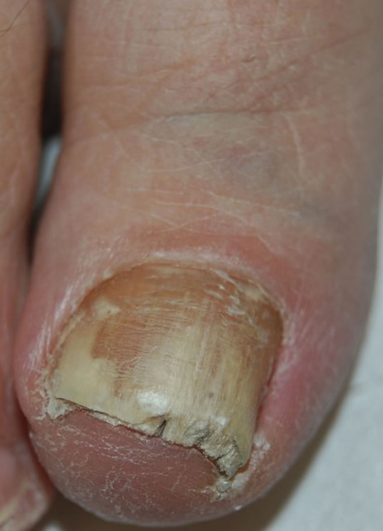
Figure 5. Distal subungual onychomycosis – with prevalent yellow discoloration
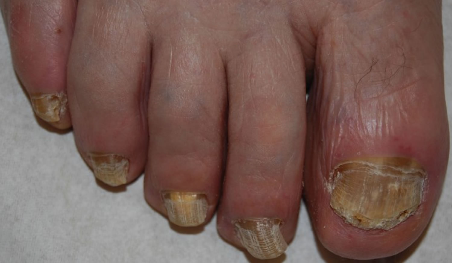
Figure 6. Distal pigmented subungual onychomycosis
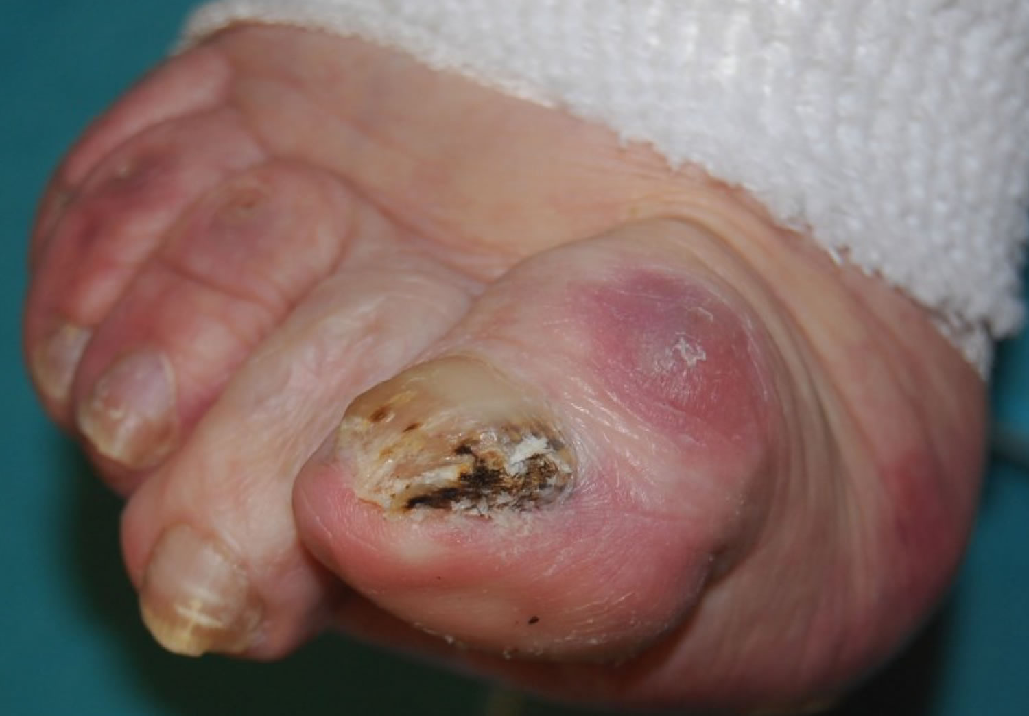
Figure 7. Onychomycosis due to molds, presenting the typical periungual inflammation.
Endonyx Onychomycosis
Endonyx onychomycosis is characterized by massive nail plate invasion in the absence of nail bed involvement. Clinically, the affected nail may show lamellar splitting and a milky white discoloration. The nail plate is firmly attached to the nail bed, and there is no nail bed hyperkeratosis or onycholysis (Figure 8) 13. This type of infection is very rare and caused by Trichophyton soudanense or Trichophyton violaceum.
Figure 8. Endonyx onychomycosis – white discoloration of the nail plate that is firmly attached to the nail bed
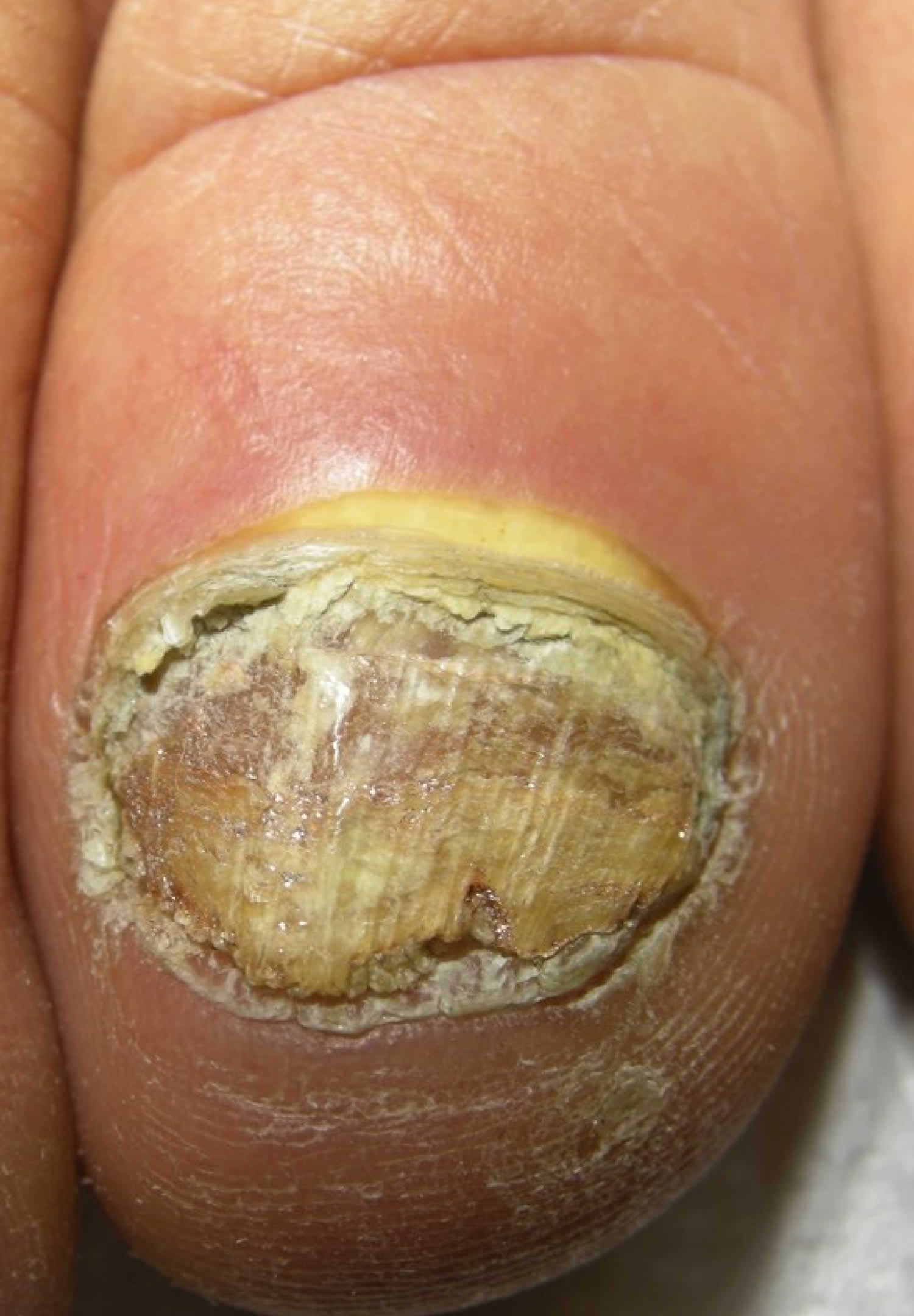
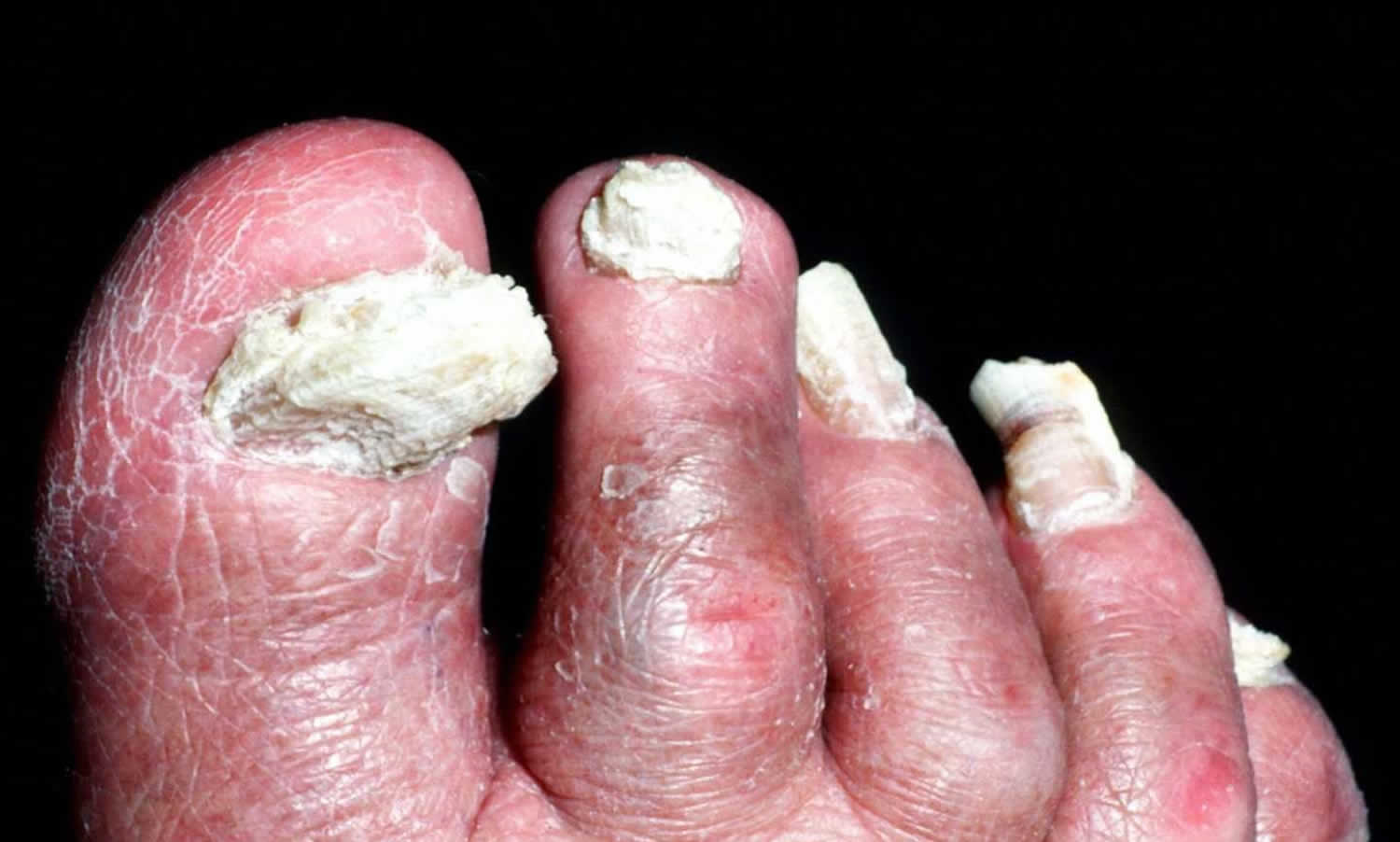
Total Dystrophic Onychomycosis
Total dystrophic onychomycosis is the most severe stage of onychomycosis, and it can result from a long-standing distal and lateral subungual onychomycosis or proximal subungual onychomycosis. The nail plate is diffusely thickened, friable and yellowish (Figures 9 and 10).
Figure 9. Total dystrophic onychomycosis – the nail plate is completely invaded by fungi and friable
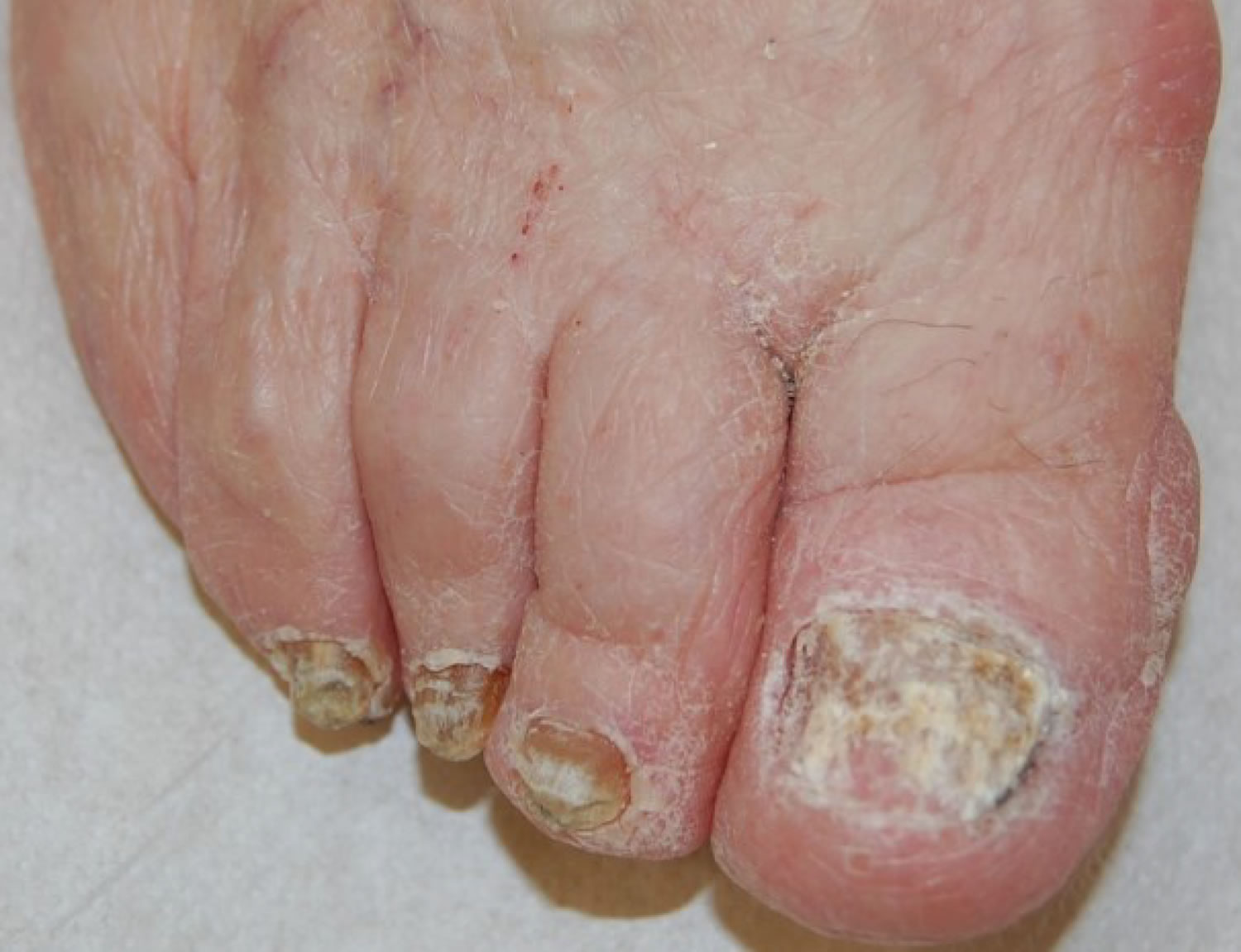
Figure 10. Onychomycosis of toenail
Onychomycosis causes
Onychomycosis is caused by various organisms, most often dermatophytes of the genus Trichophyton. Other organisms include Candida, which is more common in fingernail infections and in patients with chronic mucocutaneous candidiasis 3. Nondermatophyte molds are a less common cause in the general population. Recent studies, however, have demonstrated that they are the predominant organisms in patients with onychomycosis and human immunodeficiency virus infection (HIV) 5.
Common pathogens in onychomycosis
- Dermatophytes (80% to 90%) such as:
- Trichophyton rubrum, Trichophyton mentagrophytes, Trichophyton tonsurans and Trichophyton interdigitale. The infection is also known as tinea unguium.
- Epidermophyton floccosum
- Microsporum species
- Nondermatophytes Molds (2% to 10%): Nondermatophyte molds are the predominant organism in patients with human immunodeficiency virus infection.
- Acremonium species
- Alternaria species
- Aspergillus species
- Cladosporium carrionii
- Fusarium species
- Geotrichum candidum
- Lasiodiplodia theobromae
- Onychocola species
- Scopulariopsis species
- Scytalidium species
- Yeasts (2% to 11%) such as Candida albicans, Candida guilliermondii and Candida parapsilosis.
Fungal nail infection may occur in people with athlete’s foot (tinea pedis) and/or oozing infection (paronychia), caused by inflammation and infection with yeast and/or bacteria in the region where the skin of the finger meets the origin of the nail. Onychomycosis often spreads from athlete’s foot, a superficial infection of the skin of the feet that frequently occurs in the setting of sweaty feet.
For most people, a manicure or pedicure is a chance to pamper oneself. Unfortunately, a trip to the nail salon is not without its dangers, such as the risk of infection or damage to the nails. Here are some things you should be aware of before you treat yourself.
Onychomycosis can occur from inoculation of the nails by instruments contaminated with fungus. While it is standard procedure for nail salons to disinfect their instruments with antiseptics that kill fungi, the safest way to avoid any risk of this infection is to bring your own manicure-pedicure kit with you.
Infection of the nail fold, known as paronychia, from bacteria and/or yeast is also fairly common. Getting pedicures and manicures increases the risk of this type of infection due to removal of the cuticle, which protects the nail fold from invasion by microorganisms.
Less commonly, skin infections can occur from contaminated water in whirlpool footbaths given before pedicures. These infections usually result in folliculitis, or inflammation around the hair follicles, on the lower legs. Some of these bacteria are common organisms, such as Staphylococcal and Pseudomonas species, and respond well to standard antibiotic therapy. In unusual cases, infection with organisms known as atypical mycobacteria can occur. This usually results in more severe infection, with furuncles, or “boils,” developing on the legs in the areas exposed to the foot baths. Atypical mycobacteria do not respond well to standard antibiotics given for skin infections. Infection with atypical mycobacteria is extremely uncommon, but it can be caused by poor and improper cleaning of the whirlpool filters.
Onychomycosis prevention
Onychomycosis develop when your feet are constantly warm and damp. You’re more likely to get an infection if you wear trainers for a long time and have hot, sweaty feet.
To prevent fungal nail infections:
DO
- treat athlete’s foot as soon as possible to avoid it spreading to nails
- keep your feet clean and dry
- wear clean socks every day
- wear flip flops in showers at the gym or pool
- throw out old shoes
DON’T
- wear shoes that make your feet hot and sweaty
- share towels
- wear other people’s shoes
- share nail clippers or scissors
Preventing onychomycosis reinfection
Unfortunately, curing onychomycosis does not confer immunity. You may become reinfected at any time. The most common scenario is for the fungus to infect the skin first and then invade the nail. Thus, keeping both the skin and the nails clear of potential sources of infection is key.
Remember that nail fungus may reside in your shoes, carpet, bathroom, swimming pool, locker room, family, friends, etc. Therefore, the following are suggested:
- Don’t share nail clippers, nail instruments, shoes, etc. with others.
- Don’t cut your normal nails with the same clippers used to cut abnormal nails.
- Avoid pedicures.
- Don’t pick your toenails or scratch your feet with your finger nails.
- Use an antifungal foot powder in your shoes frequently. Alternatively, you may apply an anti-fungal cream to your feet and toes 1-2 per week.
- Family members and other close contacts should be treated if they have fungus.
- Wear flip flops in public showers, locker rooms, etc.
- When trying on new shoes, wear socks.
Onychomycosis diagnosis
Accurate diagnosis is crucial for successful treatment and requires identification of physical changes and positive laboratory analysis. Only 50% of nail problems are caused by onychomycosis 14 and clinical diagnosis by physical examination alone can be inaccurate. Psoriasis, chronic nail trauma, and other causes must also be considered. The differential diagnosis of nail fungus infection is presented in Table 1.
Table 1. Common conditions that can mimic onychomycosis
| Condition | Features |
|---|---|
Infections | |
Chronic paronychia | Chronic inflammation of the proximal paronychium; cross-striations of the nail; Streptococcus, Staphylococcus, or Candida found on smear and culture; common in children |
Viral warts | Localized in nail folds and subungual tissue; longitudinal depressed grooves in the nail plate |
Skin disorders | |
Chronic dermatitis | Subungual dermatitis, hyperkeratosis, Beau lines, and pitting; thickened nail with corrugated surface |
Lichen planus | Longitudinal grooves and fissures; usually affects fingernails |
Psoriasis | Nail pitting, splinter hemorrhages, “oil staining,” yellow-gray or silvery white nails (eFigure B) |
Twenty-nail dystrophy | Dystrophy of all 20 nails; usually resolves in childhood; associated with the lesions of lichen planus (eFigure C) |
Trauma | |
Footwear | Oncholysis, ingrown toenails, subungual keratosis, nail plate discoloration and irregularities; caused by friction against the shoe |
Manipulation (e.g., manicures, pedicures, rubbing) | Horizontal parallel nail plate grooves, inflammation from Staphylococcus aureus or Pseudomonas infection (eFigure D) |
Tumors | |
Bowen disease | Squamous cell carcinoma; bleeding, pain, nail deformity, and nail discoloration |
Fibroma | Oval or spherical, white or yellow nodule; causes tunnel-like melanonychia; fibrous dermatofibroma or periungual fibroma |
Melanoma | Brown-yellow nail with dark pigment extending into the periungual skin folds; poor prognosis |
Nail clippings should be taken from crumbling tissue at the end of the infected nail. The discolored surface of the nails can be scraped off. The debris can be scooped out from under the nail.
Previous treatment can reduce the chance of growing the fungus successfully in culture so it is best to take the clippings before any treatment is commenced:
- To confirm the diagnosis – antifungal treatment will not be successful if there is another explanation for the nail condition.
- To identify the responsible organism. Molds and yeasts may require different treatment from dermatophyte fungi.
- Treatment may be required for a prolonged period and is expensive. Partially treated infection may be impossible to prove for many months as antifungal drugs can be detected even a year later.
A nail biopsy may also reveal characteristic histopathological features of onychomycosis.
Onychomycosis treatment
Fungal nail infections can be difficult to treat. Talk with your doctor if self-care strategies and over-the-counter (nonprescription) products haven’t helped. Treatment depends on the severity of your condition and the type of fungus causing it. It can take months to see results. And even if your nail condition improves, repeat infections are common (see preventing onychomycosis reinfection above).
Treatment varies depending on the severity of nail changes, the organism involved, and concerns about adverse effects and drug interactions. Treatments also have varying effectiveness, based on cure parameters that are defined differently among studies. Mycotic cure denotes that no organism is identified on microscopy and culture. Clinical cure refers to improvement in the appearance of the nail, often defined as a normal appearance in 80% to 100% of the nail. It is a subjective measure that is difficult to compare across studies 16. Complete cure indicates that mycotic and clinical cure have been achieved.
Fingernail onychomycosis are usually cured more quickly and effectively than toenail onychomycosis.
Mild infections affecting less than 50% of one or two nails may respond to topical antifungal medications but cure usually requires an oral antifungal medication for several months. Combined topical and oral treatment is probably the most effective regime.
In the majority of the cases, patients present with a distal and lateral subungual onychomycosis due to dermatophytes involving the distal part of one or two great toenails, and the treatment of choice is topical application of antifungals, possibly associated with periodic removal of the affected nail plate. For distal and lateral subungual onychomycosis extending to the proximal nail, proximal subungual onychomycosis due to dermatophytes and deeply infiltrating white superficial onychomycosis some experts recommend systemic treatment with fluconazole, itraconazole or terbinafine. Further studies on lasers and photodynamic therapy are needed before use can be standardized.
Nonprescription agents have also been used for treatment of onychomycosis (Table 2). These therapies have been evaluated in only a small number of studies involving few patients. Topical mentholated ointment (Vicks Vaporub) was used in a small study involving 18 patients 17. After 48 weeks, 28% had mycotic and clinical cure, 56% had partial clearance, and 17% had no improvement. Tea tree oil (Melaleuca alternifolia) has been evaluated in two studies. Although one trial was favorable, combined data from both studies did not demonstrate significant benefit 18. Snakeroot extract (Ageratina pichinchensis) is an antifungal derived from plants of the sunflower family. It was studied in a randomized trial involving 96 patients who applied the extract or ciclopirox for six months to nails with confirmed infections 19. Mycotic cure occurred in 59% of patients receiving the extract and in 64% of those receiving ciclopirox. Clinical cure occurred in 71% and 81% of patients, respectively. Differences between the two treatments were not statistically significant. A small study showed that a combination of cyanoacrylate, undecylenic acid, and hydroquinone (marketed as Renewed Nail) demonstrated mycotic cure in 78 of 154 participants (50%) 20.
Table 2. Nonprescription Onychomycosis Treatments
| Agent | Administration | Clinical cure rate (%) | Mycotic cure rate (%) | Comments |
|---|---|---|---|---|
Ageratina pichinchensis (snakeroot) extract | Apply every third day for the first month, twice per week for the second month, then once per week for the third month | 71% | 59% | Study of 110 patients; therapeutic effectiveness was similar to that in the control group, which used ciclopirox |
Cyanoacrylate, undecylenic acid, and hydroquinone (Renewed Nail) | Soak and debride affected nails, then apply solution every two weeks for three to four visits; patients may also apply at home | NA | 50 to 65% (mild to moderate cases) | Study of 154 patients with cure rates reported after three months |
35% (severe cases) | ||||
Dual-wavelength near-infrared laser (Noveon) | Treatment on days 1, 14, 42, and 120 | Mild cases: 65% (3 mm of nail clearance) 26% (4 mm of nail clearance) | 30% | Toenails were evaluated on day 180 |
Moderate to severe cases: 63% (3 mm of nail clearance) | ||||
Melaleuca alternifolia (tea tree) oil | Apply twice per day | NA | NA | Cochrane review found no evidence of benefit |
Mentholated ointment (Vicks Vaporub) | Apply small amount with cotton swab daily | 28 | 28% | Pilot study of 18 patients; 56% had partial clearance, and 17% had no clearance |
Neodymium: yttrium-aluminum-garnet laser (Patholase Pinpointe) | One to three sessions four to six weeks apart | NA | 61 (complete cure) | Study of 37 toenails with onychomycosis |
19 (significant improvement) | ||||
11 (moderate improvement) |
NA = not available.
Onychomycosis medication
Antifungals from the azole and allylamine classes are the most widely used oral medications for the treatment of onychomycosis. The azole class includes itraconazole (Sporanox), fluconazole (Diflucan), and ketoconazole; however, ketoconazole is rarely prescribed because of drug interactions and hepatotoxicity. The allylamine class is represented by terbinafine (Lamisil). These medications and their dosing regimens are shown in Table 3.
Table 3. Commonly prescribed medications for treating onychomycosis in adults
| Medication | Dosing | Cure rates (%) | Organisms targeted | Potential adverse effects | Potential drug interactions* | FDA pregnancy category | Estimated monthly cost† | Comments | |
|---|---|---|---|---|---|---|---|---|---|
| Clinical | Mycotic | ||||||||
Ciclopirox 8% solution (nail lacquer) | Apply once daily to affected nails and to the underside of the nail | 6 to 9% | 29 to 36% (77 when used in combination with debridement) | Candida species, dermatophytes | Periungual erythema, erythema of the proximal nail fold, burning sensation, nail shape changes, ingrown toenails, nail discoloration | — | B | $11 for 3.3-mL bottle | Indicated for use in immunocompetent patients with mild to moderate onychomycosis without lunular involvement; patients should not bathe for eight hours after applying nail lacquer; lacquer should be removed once per week, and as much of the damaged nail as possible should be removed using scissors, nail clippers, or a nail file |
Fluconazole (Diflucan) | 100 to 300 mg orally every week for three to six months (fingernails) or six to 12 months (toenails) | 41% | 48% | Candida species | Nausea, vomiting, abdominal pain, diarrhea, headache, rash | Benzodiazepines, calcium channel blockers, statins | C | $13 for 30 100-mg tablets ($492 brand) | Not FDA approved for treatment of onychomycosis in children or adults; prescribing guidelines recommend periodic monitoring of liver function, renal function, and potassium levels; use with caution in breastfeeding women and in patients with hepatic or renal disease or porphyria |
Itraconazole (Sporanox) | Pulse dosing: 200 mg orally two times per day for one week per month, for two months (fingernails) or three months (toenails) Continuous dosing: 200 mg orally once per day for six weeks (fingernails) or 12 weeks (toenails) | 70% | 63% (pulse dosing) 69% (continuous dosing) | Candida species, dermatophytes, nondermatophyte molds, Aspergillus species | Nausea, vomiting, hypokalemia, elevated transaminase and triglyceride levels, rash | Benzodiazepines, calcium channel blockers, proton pump inhibitors, statins, warfarin (Coumadin), zolpidem (Ambien) | C | $195 for 30 100-mg capsules ($523 brand) | Liver function should be monitored in patients with preexisting hepatic dysfunction, and in all patients being treated for longer than one month; serum drug levels should be monitored because of erratic bioavailability with capsule formulation; renal function should be monitored; use with caution in breastfeeding women and in patients with hepatic or renal disease or porphyria; contraindicated in patients with ventricular dysfunction or congestive heart failure |
Terbinafine (Lamisil) | 250 mg orally once per day for six weeks (fingernails) or 12 weeks (toenails) | 66% | 76% | Some yeasts, dermatophytes, nondermatophyte molds | Gastrointestinal upset, rash, headache | Antiarrhythmic agents, beta blockers, selective serotonin reuptake inhibitors, tricyclic antidepressants, warfarin | C | $4 for 30 250-mg tablets ($607 brand) | Liver transaminase levels should be checked before therapy is started; if treatment continues beyond six weeks, complete blood count and liver function testing should be performed; use with caution in breastfeeding women and in patients with hepatic or renal disease, psoriasis, or porphyria |
[Source 8]
Oral antifungal medications
A meta-analysis of treatments for toenail onychomycosis determined that mycotic cure rates were 76% for terbinafine, 63% for itraconazole with pulse dosing, 59% for itraconazole with continuous dosing, and 48% for fluconazole 21. Clinical cure rates were 66% for terbinafine, 70% for itraconazole with pulse dosing, 70% for itraconazole with continuous dosing, and 41% for fluconazole. Common adverse effects included headache, gastrointestinal problems, and rash; these drugs also have been associated with Stevens-Johnson syndrome, long QT interval syndrome, and ventricular dysfunction. The use of these agents is discouraged in patients with liver, renal, or heart disease, and in those receiving medications with which there may be significant drug-drug interactions 22. Liver function studies are recommended before beginning treatment and after one month of therapy. A meta-analysis concluded that the risk of asymptomatic elevation of transaminase levels in immunocompetent patients receiving oral antifungal agents was 2%, and that the risk of elevations requiring termination of therapy was 1% 23. Although these medications are not approved for use in children, they have been used in children with positive results 18.
Onychomycosis topical treatment
Several topical agents are used for the treatment of onychomycosis. These agents have few contraindications and no drug-drug interactions.
Ciclopirox 8% nail lacquer is the only topical prescription medication available in the United States for the treatment of onychomycosis. It is a synthetic hydroxypyridine antifungal formulated as a nail lacquer. Adverse effects include burning, itching, and stinging at the application site 24. It may be used in patients who cannot take oral antifungals and in those with less than 50% of the distal nail affected and no lunular involvement 25. It has been used in children, although it is not approved for use in patients younger than 12 years 18. When used alone, ciclopirox has a mycotic cure rate of 29% to 36%, and a clinical cure rate of 6% to 9% 25. A Cochrane review noted that the treatment failure rate was 61% to 64% after 48 weeks of use 25.
Ciclopirox has also been used in combination with oral agents to improve effectiveness. In one comparative study, a combination of ciclopirox and oral terbinafine had a mycotic cure rate of 88% and a complete cure rate of 68%, whereas terbinafine alone had a mycotic cure rate of 65% and a complete cure rate of 50% 26.
Another possible option is Amorolfine 5% nail lacquer that is is applied once a week 27. Amorolfine has fungistatic and fungicidal properties against dermatophytes, non-dermatophytes molds and yeast 28. According to Gupta et al. 29, amorolfine 5% nail lacquer is recommended for onychomycosis without matrix involvement and mild cases of distal and lateral subungual onychomycosis that affect up to two nails.
Efinaconazole 10% solution and tavaborole 5% solution are new topical antifungals for the treatment of dermatophyte-induced onychomycosis. Efinaconazole 10% nail solution is a promising drug, approved by the FDA in June 2014, for toenail onychomycosis 30. It is a new triazole antifungal developed for topical treatment of mild to moderate distal and lateral subungual onychomycosis, applied once daily without nail debridement. Cure rates are comparable to those seen with oral itraconazole 31. A recent study 32 evaluated the efficacy of this nail lacquer on 1655 patients with onychomycosis for a period of 52 weeks, finding that efinaconazole was more effective at treating the early stage of the disease.
Tavaborole is formulated as a lightweight, water-soluble topical nail lacquer for the treatment of toenail onychomycosis 33: it has received its first global approval for this indication in the U.S. 34. Tavaborole 5% solution demonstrated efficacy and safety in phase 2 of clinical studies 35, but results from completed phase 3 studies are needed to provide additional evidences.
Terbinafine nail solution and a terbinafine spray, labeled TDT 067, may be good treatment alternatives in the future 36.
Your doctor may prescribe antifungal drugs that you take orally or apply to the nail. In some situations, it helps to combine oral and topical antifungal therapies.
- Oral antifungal drugs. These drugs are often the first choice because they clear the infection more quickly than do topical drugs. Options include terbinafine (Lamisil) and itraconazole (Sporanox). These drugs help a new nail grow free of infection, slowly replacing the infected part. You typically take this type of drug for six to 12 weeks. But you won’t see the end result of treatment until the nail grows back completely. It may take four months or longer to eliminate an infection. Treatment success rates with these drugs appear to be lower in adults over age 65.
- Oral antifungal drugs may cause side effects ranging from skin rash to liver damage. You may need occasional blood tests to check on how you’re doing with these types of drugs. Doctors may not recommend them for people with liver disease or congestive heart failure or those taking certain medications.
- Medicated nail polish. Your doctor may prescribe an antifungal nail polish called ciclopirox (Penlac). You paint it on your infected nails and surrounding skin once a day. After seven days, you wipe the piled-on layers clean with alcohol and begin fresh applications. You may need to use this type of nail polish daily for almost a year (9–12 months).
- Medicated nail cream. Your doctor may prescribe an antifungal cream, which you rub into your infected nails after soaking. These creams may work better if you first thin the nails. This helps the medication get through the hard nail surface to the underlying fungus. To thin nails, you apply a nonprescription lotion containing urea. Or your doctor may thin the surface of the nail (debride) with a file or other tool.
Physical treatments
Recently, non-drug treatment has been developed to treat onychomycosis thus avoiding the side effects and risks of oral antifungal drugs.
Nail trimming and debridement are often performed concomitantly with other treatments and appear to offer benefit. Study groups that received nail debridement with oral terbinafine had higher clinical cure rates than those who received oral terbinafine alone 37. When debridement was performed with concurrent administration of ciclopirox, the mycotic cure rate was 77%, higher than that for ciclopirox alone 38. Improvement in nail appearance was reported, but clinical cure rates were not.
Lasers emitting infrared radiation are thought to kill fungi by the production of heat within the infected tissue. Laser treatment is reported to safely eradicate nail fungi with one to three, almost painless, sessions. Several lasers have been approved for this purpose by the FDA and other regulatory authorities. However, high-quality studies of efficacy are lacking and existing studies indicate that laser treatment is less medically effective than topical or oral antifungal agents.
- Nd:YAG continuous, long or short-pulsed lasers
- Ti:Sapphire modelocked laser
- Diode laser
Photodynamic therapy using application of 5-aminolevulinic acid, methyl aminolevulinate, phenothiazine dyes (methylene blue and toluidine blue) or porphyrins, followed by exposure to red light has also been reported to be successful in small numbers of patients, whose nails were presoftened or evulsed using urea ointment for a week or so. The optimal light source and number/frequency of treatments are not established yet, so further clinical trials are needed to assess a standardized method. Photodynamic therapy using photosensitizing drugs and light to destroy fungal cells has shown some success in the treatment of onychomycosis, but further evaluation is needed 39. A recent review 40 collected a total of six articles regarding the use of photodynamic therapy in onychomycosis in vivo, but there are only case reports with small numbers of patients, except for two clinical trials. This study suggests that a previous nail abrasion or maceration (for example, with 20% urea ointment in occlusion) is needed prior to photosensitizer application. A limit of photodynamic therapy is the high number of sessions: generally three to 12 are requested. The numbers of sessions could be reduced by increasing the amount of irradiation, but with more adverse side effects, like transient pain and burning.
Iontophoresis and ultrasound are under investigation as devices used to enhance the delivery of antifungal drugs to the nail plate.
Although they are expensive, laser and photodynamic therapies have become popular based on the success of in-vitro studies. Several neodymium:yttrium-aluminum-garnet (Nd:YAG) laser therapies have been approved by the U.S. Food and Drug Administration for treatment of onychomycosis 41. The Pinpointe Foot-laser, Cutera GenesisPlus laser, and Cooltouch Varia laser are short-pulse laser systems, whereas the Light Age Q-Clear laser is a Q-switched laser. However, there are only limited data about the use of these therapies in patients. In one study, Nd:YAG laser light was used to treat 37 nails, with one to three treatments given four to eight weeks apart. At 16 weeks, 61% were completely cured, 19% had significant improvement in the nail appearance, and 11% had moderate improvement in the nail appearance 42.
Another laser treatment, the dual-wavelength near-infrared laser (Noveon), is approved for dermatologic use, but not specifically for treatment of onychomycosis 43. This treatment was used on 26 nails on days 1, 14, 42, and 120. After 180 days, 91% of nails with mild infection showed clinical improvement (3 to 4 mm of the nail free of clinical infection); however, only 30% had mycotic cure 44.
A complete systemic review 45 investigated the use of lasers on onychomycosis, including a total of 12 published papers: two randomized controlled trials; four comparative design studies (with no placebo/control group); and the others were case series, investigating in the majority of cases (10/12) the 1064-nm neodymium laser.
The author concluded that there is no consensus on laser effectiveness, due to the heterogeneity of the study designs (the definition of “cure”, duration of the study, type of onychomycosis). To date, there were no studies comparing laser with traditional therapies for onychomycosis; more information is required for a better understanding of the efficacy of this treatment.
Treatment Failure
Despite the number of available treatments, not all patients with onychomycosis are cured. Numerous factors have been cited to explain the lack of response to therapy, such as nonadherence to treatment, incorrect diagnosis, or advanced disease. Factors contributing to poor response or nonresponse to treatment are listed in Table 4.
For those who appear to be cured, recurrent infection is a risk, with a number of factors increasing the chance of recurrence. Risk factors include concomitant disease, genetic factors, immunosuppression, incorrect dosing or duration of treatment, moisture, occlusive footwear, older age, poor hygiene, tinea pedis, and trauma 46. Recurrence can be caused by lack of mycotic cure or reinfection, and the reported rate of clinical recurrence of onychomycosis ranges from 10% to 53%, regardless of the treatment method used 47. Many patients tire of continued unsuccessful treatments or recurrences, and ultimately elect to undergo permanent nail removal.
Table 4. Risk Factors for Poor Response or Nonresponse to Onychomycosis treatment
| Risk factor | Example |
|---|---|
Diagnostic problem | Another cause of nail dystrophy; mixed disease (e.g., onychomycosis and psoriasis) |
Fungal problem | Infection with drug-resistant or multiple organisms |
Nail condition that is difficult to treat | Dermatophytoma (i.e., a mass of hyphae and necrotic keratin below the nail plate); involvement of lateral aspect of the nail; more than 50% of nail affected; “spikes” extending from distal to proximal nail; subungual hyperkeratosis greater than 2 mm |
Patient characteristic or condition | Older age; diabetes mellitus; immunosuppression; impaired peripheral circulation |
Problem with medication adherence | Early termination of therapy; incorrect dosing; missed doses |
Surgery
Your doctor might suggest temporary removal of the nail so that he or she can apply the antifungal drug directly to the infection under the nail.
In stubborn (refractory) fungal nail infections that don’t respond to medicines. Your doctor might suggest permanent nail removal if the infection is severe or extremely painful.
Surgical removal of part of the nail or the entire nail, removing the nail by applying a chemical, or thinning the nail by applying 40% urea ointment may be used, in addition topical or oral antifungal agents.
References- Elewski BE. Onychomycosis: treatment, quality of life, and economic issues. Am J Clin Dermatol 2000;1:19-26.
- Kaur R, Kashyap B, Bhalla P. Onychomycosis- epidemiology, diagnosis, and management. Indian J Med Microbiol 2008;26:108-116.
- Thomas J, Jacobson GA, Narkowicz CK, Peterson GM, Burnet H, Sharpe C. Toenail onychomycosis: an important global disease burden. J Clin Pharm Ther. 2010;35(5):497–519.
- Mayser P, Freund V, Budihardja D. Toenail onychomycosis in diabetic patients: issues and management. Am J Clin Dermatol. 2009;10(4):211–220.
- Surjushe A, Kamath R, Oberai C, et al. A clinical and mycological study of onychomycosis in HIV infection. Indian J Dermatol Venereol Leprol. 2007;73(6):397–401.
- Roujeau JC, Sigurgeirsson B, Korting HC, Kerl H, Paul C. Chronic dermatomycoses of the foot as risk factors for acute bacterial cellulitis of the leg: a case-control study. Dermatology. 2004;209(4):301–307.
- Boyko EJ, Ahroni JH, Cohen V, Nelson KM, Heagerty PJ. Prediction of diabetic foot ulcer occurrence using commonly available clinical information: the Seattle Diabetic Foot Study. Diabetes Care. 2006;29(6):1202–1207.
- Onychomycosis: Current Trends in Diagnosis and Treatment. Am Fam Physician. 2013 Dec 1;88(11):762-770. https://www.aafp.org/afp/2013/1201/p762.html
- Baran R, Kaoukhov A. Topical antifungal drugs for the treatment of onychomycosis: an overview of current strategies for monotherapy and combination therapy. J Eur Acad Dermatol Venereol. 2005;19(1):21–29.
- Piraccini, B.M.; Tosti, A. White superficial onychomycosis: Epidemiological, clinical, and pathological study of 79 patients. Arch. Dermatol. 2004, 140, 696–701.
- Pichardo-Geisinger, R.; Mora, D.C.; Newman, J.C.; Arcury, T.A.; Feldman, S.R.; Quandt, S.A. Comorbidity of tinea pedis and onychomycosis and evaluation of risk factors in Latino immigrant poultry processing and other manual laborers. South. Med. J. 2014, 107, 374–349.
- Finch, J.; Arenas, R.; Baran, R. Fungal melanonychia. J. Am. Acad. Dermatol. 2012, 66, 830–841.
- Tosti, A.; Baran, R.; Piraccini, B.M.; Fanti, P.A. “Endonyx” onychomycosis: A new modality of nail invasion by dermatophytes. Acta Derm. Venereol. 1999, 79, 52–53.
- Faergemann J, Baran R. Epidemiology, clinical presentation and diagnosis of onychomycosis. Br J Dermatol. 2003;149(suppl 65):1–4.
- Allevato MA. Diseases mimicking onychomycosis. Clin Dermatol. 2010;28(2):164–177.
- Scher RK, Tavakkol A, Sigurgeirsson B, et al. Onychomycosis: diagnosis and definition of cure. J Am Acad Dermatol. 2007;56(6):939–944.
- Landsman AS, Robbins AH, Angelini PF, et al. Treatment of mild, moderate, and severe onychomycosis using 870- and 930-nm light exposure. J Am Podiatr Med Assoc. 2010;100(3):166–177.
- Gupta AK, Skinner AR. Onychomycosis in children: a brief overview with treatment strategies. Pediatr Dermatol. 2004;21(1):74–79.
- Romero-Cerecero O, Zamilpa A, Jiménez-Ferrer JE, Rojas-Bribiesca G, Román-Ramos R, Tortoriello J. Double-blind clinical trial for evaluating the effectiveness and tolerability of Ageratina pichinchensis extract on patients with mild to moderate onychomycosis. A comparative study with ciclopirox [published correction appears in Planta Med. 2008;74(14):1767]. Planta Med. 2008;74(12):1430–1435.
- Rehder P, Nguyen TT. A new concept in the topical treatment of onychomycosis with cyanoacrylate, undecylenic acid, and hydroquinone. Foot Ankle Spec. 2008;1(2):93–96.
- Gupta AK, Ryder JE, Johnson AM. Cumulative meta-analysis of systemic antifungal agents for the treatment of onychomycosis. Br J Dermatol. 2004;150(3):537–544.
- Antifungal drugs. Treat Guidel Med Lett. 2009;7(88):95–102.
- Chang CH, Young-Xu Y, Kurth T, Orav JE, Chan AK. The safety of oral antifungal treatments for superficial dermatophytosis and onychomycosis: a meta-analysis. Am J Med. 2007;120(9):791–798.
- Rotta I, Sanchez A, Gonçalves PR, Otuki MF, Correr CJ. Efficacy and safety of topical antifungals in the treatment of dermatomycosis: a systematic review. Br J Dermatol. 2012;166(5):927–933.
- Gupta AK, Fleckman P, Baran R. Ciclopirox nail lacquer topical solution 8% in the treatment of toenail onychomycosis. J Am Acad Dermatol. 2000;43(4 suppl):S70–S80.
- Avner S, Nir N, Henri T. Combination of oral terbinafine and topical ciclopirox compared to oral terbinafine for the treatment of onychomycosis. J Dermatolog Treat. 2005;16(5–6):327–330.
- Gupta, A.K.; Daigle, D.; Foley, K.A. Topical therapy for toenail onychomycosis: An evidence-based review. Am. J. Clin. Dermatol. 2014, 15, 489–502.
- Gupta, A.K.; Ryder, J.E.; Baran, R. The use of topical therapies to treat onychomycosis. Dermatol. Clin. 2003, 21, 481–489.
- Gupta, A.K.; Paquet, M.; Simpson, F.C. Therapies for the treatment of onychomycosis. Clin. Dermatol. 2013, 31, 544–554.
- Elewski, B.E.; Rich, P.; Pollak, R.; Pariser, D.M.; Watanabe, S.; Senda, H.; Ieda, C.; Smith, K.; Pillai, R.; Ramakrishna, T.; et al. Efinaconazole 10% solution in the treatment of toenail onychomycosis: Two phase III multicenter, randomized, double-blind studies. J. Am. Acad. Dermatol. 2013, 68, 600–608.
- Tosti, A. Efinaconazole solution 10%: Topical antifungal therapy for toenail onychomycosis. Cutis 2013, 92, 203–208.
- Rich, P. Efinaconazole topical solution, 10%: The benefits of treating onychomycosis early. J. Drugs Dermatol. 2015, 14, 58–62.
- Elewski, B.E.; Tosti, A. Tavaborole for the treatment of onychomycosis. Expert Opin. Pharmacother. 2014, 15, 1439–1448.
- Markham, A. Tavaborole: First global approval. Drugs 2014, 74, 1555–1558.
- Toledo-Bahena, M.E.; Bucko, A.; Ocampo-Candiani, J.; Herz-Ruelas, M.E.; Jones, T.M.; Jarratt, M.T.; Pollak, R.A.; Zane, L.T. The efficacy and safety of tavaborole, a novel, boron-based pharmaceutical agent: Phase 2 studies conducted for the topical treatment of toenail onychomycosis. J. Drugs Dermatol. 2014, 13, 1124–1132.
- Elewski, B.E.; Ghannoum, M.A.; Mayser, P.; Gupta, A.K.; Korting, H.C.; Shouey, R.J.; Baker, D.R.; Rich, P.A.; Ling, M.; Hugot, S.; et al. Efficacy, safety and tolerability of topical terbinafine nail solution in patients with mild to moderate toenail onychomycosis: Results from three randomized studies using double-blind vehicle controlled and open-label active controlled designs. J. Eur. Acad. Dermatol. Venereol. 2013, 27, 287–294.
- Jennings MB, Pollak R, Harkless LB, Kianifard F, Tavakkol A. Treatment of toenail onychomycosis with oral terbinafine plus aggressive debridement: IRON-CLAD, a large, randomized, open-label, multicenter trial. J Am Podiatr Med Assoc. 2006;96(6):465–473.
- Malay DS, Yi S, Borowsky P, Downey MS, Mlodzienski AJ. Efficacy of debridement alone versus debridement combined with topical antifungal nail lacquer for the treatment of pedal onychomycosis: a randomized, controlled trial. J Foot Ankle Surg. 2009;48(3):294–308.
- Sotiriou E, Koussidou-Eremonti T, Chaidemenos G, Apalla Z, Ioannides D. Photodynamic therapy for distal and lateral subungual toenail onychomycosis caused by Trichophyton rubrum: preliminary results of a single-centre open trial. Acta Derm Venereol. 2010;90(2):216–217.
- Simmons, B.J.; Griffith, R.D.; Falto-Aizpurua, L.A.; Nouri, K. An update on photodynamic therapies in the treatment of onychomycosis. J. Eur. Acad. Dermatol. Venereol. 2015.
- Gupta AK, Simpson F. Newly approved laser systems for onychomycosis. J Am Podiatr Med Assoc. 2012;102(5):428–430.
- Kimura U, Takeuchi K, Kinoshita A, Takamori K, Hiruma M, Suga Y. Treating onychomycoses of the toenail: clinical efficacy of the sub-millisecond 1,064 nm Nd:YAG laser using a 5 mm spot diameter. J Drugs Dermatol. 2012;11(4):496–504.
- 510(k) summary: Noveon (Model LS1100-01-0968) dual wavelength leser instrument. https://www.accessdata.fda.gov/cdrh_docs/pdf7/K071815.pdf
- Landsman AS, Robbins AH. Treatment of mild, moderate, and severe onychomycosis using 870- and 930-nm light exposure: some follow-up observations at 270 days. J Am Podiatr Med Assoc. 2012;102(2):169–171.
- Bristow, I.R. The effectiveness of lasers in the treatment of onychomycosis: A systematic review. J. Foot Ankle Res. 2014, 7.
- Scher RK, Baran R. Onychomycosis in clinical practice: factors contributing to recurrence. Br J Dermatol. 2003;149(suppl 65):5–9.
- Piraccini BM, Sisti A, Tosti A. Long-term follow-up of toenail onychomycosis caused by dermatophytes after successful treatment with systemic antifungal agents. J Am Acad Dermatol. 2010;62(3):411–414.


