What is porokeratosis
Porokeratosis is a group of uncommon skin conditions in which there is abnormal keratinization 1. The name porokeratosis means scaly pore, and is a misnomer as porokeratosis is not related to pores. Porokeratosis is a disorder of keratinization that presents with keratotic papules or annular plaques with a well-defined ridge-like border 2. The skin lesions that result are dry and atrophic, with a well-defined ridge-like hyperkeratotic border called a cornoid lamella (best seen on dermoscopy) 3. Porokeratosis has a distinct histologic hallmark of cornoid lamella 4, which is a column of tightly fitted parakeratotic cells in the upper epidermis 5. Porokeratosis is a precancerous lesion that can undergo malignant transformation 6. Evaluation of porokeratosis is best done with biopsy of the elevated border.
Porokeratosis can be present at birth or not develop until adult life 7, depending on the type of porokeratosis.
There are multiple different clinical variants of porokeratosis, including disseminated superficial actinic porokeratosis, classical porokeratosis of Mibelli, porokeratosis palmaris plantaris et disseminatum, and linear porokeratosis 4. Additionally, more rare variants include genitogluteal porokeratosis, facial porokeratosis, giant porokeratosis, porokeratosis ptychotropica, hypertrophic verrucous porokeratosis, eruptive pruritic papular porokeratosis, follicular porokeratosis, and reticulate porokeratosis 8. Variants can occur together 9, but rarely do 10.
While multiple therapies are described for porokeratosis, including topical, systemic, and surgical, there are no standard guidelines for treatment 11.
What are the types of porokeratosis?
Types of porokeratosis include:
- Disseminated superficial actinic porokeratosis
- Porokeratosis of Mibelli
- Linear porokeratosis
- Porokeratosis ptychotropica
- Porokeratosis of Mantoux (also known as porokeratosis palmaris et plantaris disseminata, which is a form of punctate palmoplantar keratoderma in which
- there are scaly red-brown annular patches on the palms and soles that later spread to the limbs and trunk)
- Porokeratotic eccrine ostial and dermal duct naevus
- Porokeratotic adnexal ostial nevus (PAON)
- Pruritic papular porokeratosis
- Pigmented porokeratosis
- Punctate follicular porokeratosis
Disseminated actinic porokeratosis
Disseminated superficial actinic porokeratosis is an inherited keratinisation disorder that causes discrete dry patches on the arms and legs. Disseminated superficial actinic porokeratosis is a special type of inherited ‘sunspot” 12.
Disseminated superficial actinic porokeratosis most often affects people of European descent, although it has also been reported to affect individuals of other races. It is more common in women than in men.
The average age which patients first notice Disseminated superficial actinic porokeratosis is about 35–40 years and its frequency in affected families increases steadily with age. It is rare in childhood.
Disseminated superficial actinic porokeratosis may arise in immune suppressed patients, including after organ transplantation. Its onset can also be triggered by sun exposure, phototherapy, injury, infection or systemic disease.
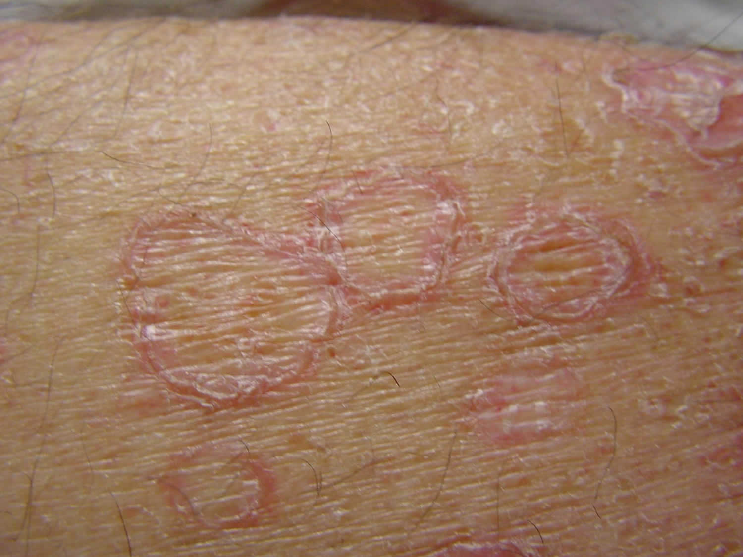
Figure 1. Disseminated superficial actinic porokeratosis

What causes disseminated superficial actinic porokeratosis?
Disseminated superficial actinic porokeratosis is due to a genetic mutation. The tendency to Disseminated superficial actinic porokeratosis is inherited as an autosomal dominant characteristic, which means on average half of the children of an affected parent will also have the tendency.
A locus on chromosome 12 was found to be responsible for Disseminated superficial actinic porokeratosis in a Chinese family 14. The causative genes in affected families have included spliceosome-associated factor 3 (SART3) and mevalonate kinase (MVK) genes.
Disseminated superficial actinic porokeratosis signs and symptoms
Disseminated superficial actinic porokeratosis mainly affects the lower arms and legs bilaterally and arises more frequently on the lower legs. There may be few or innumerable lesions. The forehead and cheeks are affected in less than 10% of individuals and Disseminated superficial actinic porokeratosis almost never occurs on the scalp, palms or soles. It tends to be more prominent in the summer and may appear less prominent in winter. New lesions have been provoked by ultraviolet light in sun lamps.
The lesions are composed of multiple irregular roundish, annular or polycyclic plaques, each of which has an elevated horny rim. The visibility of this rim is markedly accentuated by the application of artificial tanning solution (dihydroxyacetone).
The smallest Disseminated superficial actinic porokeratosis lesion is a 1–3 mm conical papule, skin coloured, brownish red or brown in colour. It is based around a hair follicle containing a keratotic (scaly) plug. Larger plaques have a sharp, slightly raised, keratotic ring, a fraction of a millimetre thick, with a diameter of 10 mm or more. The skin within the ring is thinned and mildly reddened or slightly brown, and a pale ring may be seen just within the ridge. The ridge itself is often a darker brown than the rest of the lesion. The central area is most often pale and smooth, but it may be red, scaly, dry, or have scaly follicular plugs.
Sweating is absent within the lesions. Although most often asymptomatic, sun exposure or heat may cause them to itch or sting.
Disseminated superficial actinic porokeratosis complications
Development of squamous cell carcinoma (SCC) within a Disseminated superficial actinic porokeratosis lesion is the main concern. This is uncommon (< 10% of individuals with Disseminated superficial actinic porokeratosis develop squamous cell carcinoma). However, many patients with Disseminated superficial actinic porokeratosis have had significant exposure to the sun and may also have actinic keratoses and other forms of skin cancer (particularly basal cell carcinoma). SCC presents as a solitary tender enlarging scaly or ulcerated plaque or nodule.
Disseminated superficial actinic porokeratosis diagnosis
The diagnosis of porokeratosis is usually clinical, with the help of dermoscopy. Disseminated superficial actinic porokeratosis is sometimes diagnosed by finding characteristic features on pathology, in which the scaly rim of Disseminated superficial actinic porokeratosis is described as a parakeratotic cornoid lamella. The diagnosis can also be missed on a biopsy if the specimen does not include the rim, it is poorly orientated, or the pathologist’s attention is not drawn to the horny ridge seen clinically.
Disseminated superficial actinic porokeratosis treatment
Unfortunately, in the present state of knowledge, there is no very satisfactory treatment for disseminated superficial actinic porokeratosis. Over the years the agents that have been tried include:
- Cryotherapy
- 5-Fluorouracil cream
- Imiquimod cream
- Tretinoin cream
- Ingenol mebutate gel
- Alpha hydroxy acid cream
- Calcipotriol ointment
- Diclofenac gel
- Oral acitretin
- Photodynamic therapy
- Grenz ray therapy
- Laser treatments.
To date, no treatment has proved effective long term. Most people settle for just having the larger lesions frozen lightly and returning as necessary for further treatments, using a moisturiser to reduce the dry feeling.
If the disseminated superficial actinic porokeratosis has been induced by drug-induced immune suppression, withdrawal of the drug has been reported to result in remission of disseminated superficial actinic porokeratosis.
Sun protection
Restriction of sun exposure by wearing long sleeves, skirts or slacks and using sunscreens on the legs and arms is believed to reduce or delay the development of new lesions.
Porokeratosis of Mibelli
Porokeratosis of Mibelli is the second most common form of porokeratosis 15. Porokeratosis of Mibelli most often starts during childhood but lesions may be present at birth or may first appear at puberty or later 15. Sometimes it appears during adulthood following the suppression of the immune system by certain medication or illness 16.
Porokeratosis of Mibelli affects men twice as commonly as women 15.
Figure 2. Porokeratosis of Mibelli
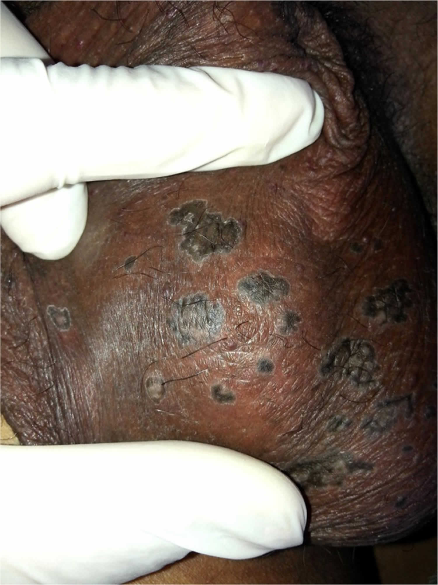
Footnote: Porokeratosis of Mibelli on the scrotum showing the peripheral ridge that appears hypopigmented and completely surrounds the pigmented annular plaques.
[Source 17 ]What causes porokeratosis of Mibelli?
The cause of porokeratosis of Mibelli is unknown. Occasionally there is a family history of porokeratosis of Mibelli or another type of porokeratosis such as linear porokeratosis or disseminated superficial actinic porokeratosis, suggesting there is a genetic predisposition to the disorder.
What are the clinical features of porokeratosis of Mibelli?
The porokeratosis lesion starts as a small, light brown, scaly papule. Several papules may join into one or more plaques with irregular boundaries. Each plaque is separated from the surrounding skin by a warty rim with well-defined thin furrows running in its centre.
Porokeratosis of Mibelli most often affects the limbs, particularly the hands and feet, lower leg, neck, shoulders, face or genitals, although any part of the body may be affected including the mucous membranes 18.
The lesions usually reach only a few centimetres in size but ‘giant porokeratomas’ can be as large as 10 to 20 cm wide 19.
Porokeratosis of Mibelli complications
Lesions may remain unchanged for many years or may slowly grow between long periods of inactivity. A skin cancer can develop within porokeratosis of Mibelli. This may be either a basal or squamous cell carcinoma and is more likely to occur in older adults.
Rarely, porokeratosis of Mibelli may involve the distal fingers or toes and this can result in damage to the nails 20.
Porokeratosis of Mibelli diagnosis
The diagnosis of porokeratosis is usually made clinically but sometimes a biopsy is needed. The pathology is very distinct for this condition.
Porokeratosis of Mibelli treatment
There is no known cure for porokeratosis of Mibelli and treatment is generally disappointing. However, the appearance may improve with the following measures:
- 5-Fluorouracil cream
- Calcipotriol cream
- Oral acitretin or isotretinoin
- Cryotherapy
- Dermabrasion
- Carbon dioxide laser ablation
Sun protection is very important as exposure to ultraviolet radiation may result in the development of skin cancer within the porokeratosis.
Porokeratosis of Mibelli prognosis
The risk of progression of porokeratosis of Mibelli to a basal or squamous cell carcinoma is about 10% 21. This presents as a changing, enlarging crusted sore or nodule. It may require a biopsy or is cut out (excision).
Linear porokeratosis
In linear porokeratosis, the lesions are arranged in a linear formation 22. Linear porokeratosis can be present at birth or not develop until adult life 22.
Linear porokeratosis causes
Like other forms of porokeratosis, the cornoid lamella in linear porokeratosis is due to an expanding proliferation of unusual keratinocytes, which is thought to be due to a genetic mutation. Genetic mutations presenting in the form of mosaicism would explain the linearity and often unilateral distribution of the lesions 23.
Occasionally there is a family history of linear porokeratosis or another kind of porokeratosis, such as disseminated superficial actinic porokeratosis, consistent with a genetic predisposition 24.
Linear porokeratosis clinical features
Linear porokeratosis presents as numerous grouped lesions, each with the characteristic ridge on its border and a central furrow.
They are arranged in one or more lines along a limb or on one side of the trunk, head and neck, following a dermatomal distribution (ie, along the pathway of a sensory nerve).
Localised linear porokeratosis is unilateral and is often confined to a single extremity. The generalised form of linear porokeratosis is rarer, and the lesions affect several extremities and the trunk 25.
Linear porokeratosis complications
The main complication of linear porokeratosis is skin cancer, which can develop within a linear porokeratosis lesion. This may be either a basal cell carcinoma or squamous cell carcinoma and is more likely to occur in older adults 26. Linear porokeratosis should be monitored for malignancy.
Linear porokeratosis diagnosis
The diagnosis of linear porokeratosis is usually made clinically (based on the appearance), but sometimes a biopsy is needed. The biopsy should include the raised edge of the lesion. The pathology of porokeratosis is very distinct, but it may be necessary to point out the clinical features for the pathologist to find a cornoid lamella within the pathological specimen.
Linear porokeratosis treatment
There is no known cure for linear porokeratosis and treatment is generally disappointing. However, the appearance may improve with the following measures:
- 5-fluorouracil cream
- Calcipotriol cream
- Oral acitretin or isotretinoin
- Cryotherapy
- Dermabrasion
- Carbon dioxide laser ablation.
Sun protection is very important as exposure to ultraviolet radiation may result in the development of skin cancer within the linear porokeratosis.
What causes porokeratosis?
The etiology of porokeratosis is unclear at this time 5. Porokeratosis usually occurs on sun-exposed skin, most commonly in the fifth decade of life 27 but can occur at any age and at similar frequencies in males and females 27. The increased occurrence of disseminated superficial actinic porokeratosis on sun-exposed skin likely indicates that ultraviolet light is a risk factor 28.
The eruptive form of porokeratosis is associated with immune suppression, transplantation patients, inflammatory states, and malignancy 29.
Porokeratosis will progress to nonmelanoma skin cancer in 6.9% to 30% of cases, most frequently squamous cell carcinoma and less frequently basal cell carcinoma 6.
While porokeratosis is usually an acquired disease, there is often a familial predisposition which could signify a hereditary component 9.
Porokeratosis pathophysiology
Porokeratosis is an entity that results from disordered progression of epidermal cells 30. The development of this entity can be related to sun exposure. In areas where there has been no or limited sun exposure, repeated minor frictional trauma due to tight clothing may cause the entity 10, as is the case in genitourinary porokeratosis.
There is reported association with overexpression of p53 6 and occasionally there can be an expansion of a clone of abnormal epidermal keratinocytes 9. Porokeratosis has the possibility of malignant transformation into squamous cell carcinoma or basal cell carcinoma 9.
Porokeratosis histopathology
The distinctive histopathologic feature of porokeratosis is a cornoid lamella 27, which is a column of tightly fitted parakeratotic cells 31. The column of parakeratosis will occur over the epidermis with an absent granular layer and dyskeratotic cells in the upper spinous zone 10. This feature is usually present at the elevated border of the lesion 10. While once thought to be pathognomonic of porokeratosis, the feature has been found in other conditions 30. Cornoid lamellation reflects a disordered progression of epidermal cells 30. In follicular porokeratosis, the cornoid lamella can involve the follicular infundibulum 32.
What are the clinical features of porokeratosis?
Each porokeratosis lesion has a characteristic ridge on its border and a central furrow. Their size, onset and distribution depends on the specific type of porokeratosis.
Porokeratosis presents as keratotic papules or annular plaques that expand centrifugally with an elevated keratotic border 10. Centrally, the lesion can appear slightly atrophic 10. The lesions can present with pruritus and may be present for several years before diagnosis 10. Disseminated nodules present as pink to brown papules and macules with raised borders 29. Porokeratosis most commonly occurs in the limbs and trunk but can occur in the trunk, face, and genitourinary region, and scrotum 32. However, porokeratosis as an entity has a broad spectrum of presentations, including large destructive lesions and involvement of burn scars 33.
Porokeratosis complications
The main complication of porokeratosis is a skin cancer, which can develop within a porokeratosis lesion. This may be either a basal cell carcinoma or squamous cell carcinoma, and is more likely to occur in older adults 26. Porokeratosis should be monitored for malignancy.
Porokeratosis diagnosis
The diagnosis of porokeratosis is usually made clinically, often with the help of dermoscopy, but sometimes a biopsy is needed. The biopsy should include the raised edge of the lesion 17. The pathology of porokeratosis is very distinct, but it may be necessary to point out the clinical features for the pathologist to find a cornoid lamella within the pathological specimen.
Dermoscopy of lesions shows central brown pigmentation with blue-gray dots surrounded by a single hypopigmented band with a white track at the periphery 17.
Porokeratosis treatment
There is no known cure for porokeratosis and treatment is generally disappointing. Multiple therapies are described for porokeratosis, including topical, systemic, and surgical. However, there have been no randomized controlled trials, so there are no international guidelines on treatment standards 34.
- Classical porokeratosis of Mibelli is most successfully treated with imiquimod cream 34.
- Linear porokeratosis responds well to topical or systemic retinoids 34.
- Disseminated porokeratosis can be treated successfully with topical vitamin D acid derivatives 34.
- Surgical interventions or cryotherapy are options for treatment in areas where topical agents are difficult to use or contraindicated 34.
- Laser therapy may be another treatment option 29.
- Topical steroids, retinoids, and topical diclofenac may provide symptomatic relief even if no lasting benefit is achieved 17.
Sun protection is important as exposure to ultraviolet radiation may result in the development of a skin cancer within the linear porokeratosis.
Porokeratosis prognosis
Porokeratosis is a premalignant condition 17; however, all types of porokeratosis can undergo malignant transformation to non-melanoma skin cancer 27 at a rate of 6.9-30% 33. Most commonly, the lesion transforms into squamous cell carcinoma. Less commonly, the lesion transforms into basal cell carcinoma. If the lesion has not undergone malignant transformation, excision is curative.
Linear porokeratosis and giant porokeratosis are the variants that are most susceptible to malignant transformation. Malignant transformation is most rare in disseminated superficial actinic porokeratosis 35.
References- Williams GM, Fillman EP. Porokeratosis. [Updated 2019 Apr 4]. In: StatPearls [Internet]. Treasure Island (FL): StatPearls Publishing; 2019 Jan-. Available from: https://www.ncbi.nlm.nih.gov/books/NBK532290
- Joshi R, Minni K. Genitogluteal porokeratosis: a clinical review. Clin Cosmet Investig Dermatol. 2018;11:219-229
- Gutierrez EL, Galarza C, Ramos W, Tello M, De Paz PC, Bobbio L, Barquinero A, Ronceros G, Ortega-Loayza AG. Facial porokeratosis: A series of six patients. Australas J Dermatol. 2010 Aug;51(3):191-4. doi: 10.1111/j.1440-0960.2009.00616.x
- Leow YH, Soon YH, Tham SN. A report of 31 cases of porokeratosis at the National Skin Centre. Ann. Acad. Med. Singap. 1996 Nov;25(6):837-41
- Sertznig P, von Felbert V, Megahed M. Porokeratosis: present concepts. J Eur Acad Dermatol Venereol. 2012 Apr;26(4):404-12
- Ahmed A, Hivnor C. A case of genital porokeratosis and review of literature. Indian J Dermatol. 2015 Mar-Apr;60(2):217
- Rahbari H, Cordero AA, Mehregan AH. Linear porokeratosis. A distinctive clinical variant of porokeratosis of Mibelli. Arch Dermatol. 1974 Apr;109(4):526-8.
- Le C, Bedocs PM. StatPearls [Internet]. StatPearls Publishing; Treasure Island (FL): Feb 13, 2019. Disseminated Superficial Actinic Porokeratosis.
- Kanitakis J. Porokeratoses: an update of clinical, aetiopathogenic and therapeutic features. Eur J Dermatol. 2014 Sep-Oct;24(5):533-44.
- Joshi R, Minni K. Genitogluteal porokeratosis: a clinical review. Clin Cosmet Investig Dermatol. 2018;11:219-229.
- Weidner T, Illing T, Miguel D, Elsner P. Treatment of Porokeratosis: A Systematic Review. Am J Clin Dermatol. 2017 Aug;18(4):435-449
- Disseminated superficial actinic porokeratosis. https://www.dermnetnz.org/topics/disseminated-superficial-actinic-porokeratosis/
- Meo, N. D, Fluehler, C., Perkan, V., & Trevisan, G. (2010). Disseminated superficial porokeratosis and pyoderma gangrenosum. Dermatology Online Journal, 16(10). Retrieved from https://escholarship.org/uc/item/5n1901d5
- Zhu T, Tian D, Zhang L, Xu X, Xia K, Hu Z, Xiong Z, Tan J. Novel mutations in mevalonate kinase cause disseminated superficial actinic porokeratosis. Br J Dermatol. 2018 Dec 30. doi: 10.1111/bjd.17596
- Sertznig P, von Felbert V, Megahed M. Porokeratosis: present concepts. J Eur Acad Dermatol Venereol 2012; 26: 404–12.
- Rodriguez EA, et al. Porokeratosis of Mibelli and HIV-infection. Int J Dermatol; 35: 402–4.
- Joshi R, Minni K. Genitogluteal porokeratosis: a clinical review. Clin Cosmet Investig Dermatol. 2018;11:219–229. Published 2018 May 1. doi:10.2147/CCID.S143085 https://www.ncbi.nlm.nih.gov/pmc/articles/PMC5936488
- Mehregan AH, Khalili H, Fazel Z. Mibelli porokeratosis of the face. A report of seven cases. J Am Acad Dermatol 1980; 3: 394–6.
- Bozdag KE, Bicakci H, Ermete M. Giant porokeratosis. Int J Dermatol 2004; 43: 518–20.
- Rajesh G, et al. Acral porokeratosis associated with anonychia. Indian J Dermatol Venereol Leprol 2018; 84: 81–82.
- Sasson M, Krain AD. Porokeratosis and cutaneous malignancy. A review. Dermatol Surg 1996; 22: 339–42.
- Rahbari H, Cordero AA, Mehregan AH. Linear porokeratosis. A distinctive clinical variant of porokeratosis of Mibelli. Arch Dermatol. 1974 Apr;109(4):526-8
- Happle R. Segmental forms of autosomal dominant skin disorders: different types of severity reflect different states of zygosity. Am J Med Genet. 1996 Dec 11;66(2):241-2.
- Kaur S, Thami GP, Mohan H, Kanwar AJ. Co-existence of variants of porokeratosis: a case report and a review of the literature. J Dermatol. 2002 May;29(5):305-9.
- Dervis E, Demirkesen C. Generalized linear porokeratosis. Int J Dermatol. 2006 Sep;45(9):1077-9.
- Sasson M, Krain AD. Porokeratosis and cutaneous malignancy. A review. Dermatol Surg. 1996 Apr;22(4):339-42.
- Leow YH, Soon YH, Tham SN. A report of 31 cases of porokeratosis at the National Skin Centre. Ann. Acad. Med. Singap. 1996 Nov;25(6):837-41.
- Neumann RA, Knobler RM, Jurecka W, Gebhart W. Disseminated superficial actinic porokeratosis: experimental induction and exacerbation of skin lesions. J. Am. Acad. Dermatol. 1989 Dec;21(6):1182-8.
- Le C, Bedocs PM. StatPearls [Internet]. StatPearls Publishing; Treasure Island (FL): Feb 13, 2019. Disseminated Superficial Actinic Porokeratosis.
- Wade TR, Ackerman AB. Cornoid lamellation. A histologic reaction pattern. Am J Dermatopathol. 1980 Spring;2(1):5-15.
- Sertznig P, von Felbert V, Megahed M. Porokeratosis: present concepts. J Eur Acad Dermatol Venereol. 2012 Apr;26(4):404-12.
- Zhao M, Sanusi T, Zhao Y, Huang C, Chen S. Porokeratosis with follicular involvement: report of three cases and review of literatures. Int J Clin Exp Pathol. 2015;8(4):4248-52.
- Ahmed A, Hivnor C. A case of genital porokeratosis and review of literature. Indian J Dermatol. 2015 Mar-Apr;60(2):217.
- Weidner T, Illing T, Miguel D, Elsner P. Treatment of Porokeratosis: A Systematic Review. Am J Clin Dermatol. 2017 Aug;18(4):435-449.
- Silver SG, Crawford RI. Fatal squamous cell carcinoma arising from transplant-associated porokeratosis. J. Am. Acad. Dermatol. 2003 Nov;49(5):931-3.


