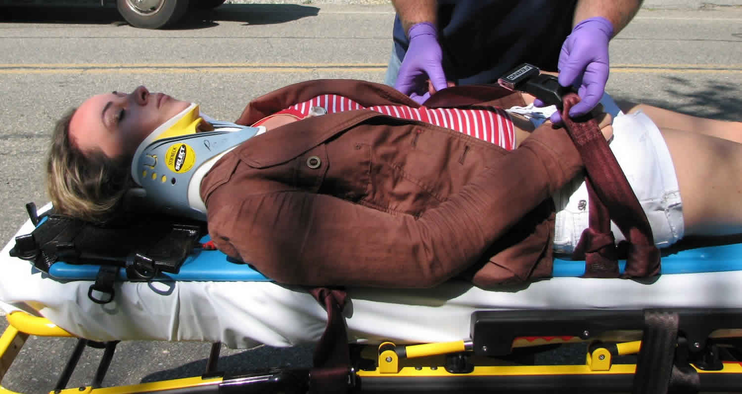
Spinal precautions
Spinal precautions also known as spinal immobilization and spinal motion restriction, are efforts to prevent movement of the spine in those with a risk of a spine injury. This is done as an effort to prevent injury to the spinal cord 1. Spinal precautions include head holding, application of a cervical collar, patients nursed in neutral alignment on an approved mattress and log rolling for all care. These precautions should be in place until clinical and or radiological examination has been performed to establish that the spine has been ‘cleared’ or a management plan has been made 2. Spinal precautions involve the same care and attention to spinal protection as spinal immobilization, with the only exception being the judicious use of the backboard or similarly rigid devices during transport. Because “spinal immobilization” and “backboarding” have become synonyms, changes in emergency medical services (EMS) protocols to adopt “spinal precautions” by decreasing or eliminating the backboard as a spinal protection device during transport represent a significant change in emergency medical services (EMS) practice and culture within the United States. Other modern EMS systems have already made significant change in practice. In much of Australia, the backboard is used only as an extrication device with use during transport being discouraged or prohibited 3.
It is estimated that 2% of people with blunt trauma will have a spinal cord injury, and these rates are higher in the setting of severe closed head injury 4. Spine injury (cord and/or column) must be considered a possibility in any patient with significant trauma to the head or torso with the most common mechanism of injuries being motor vehicle accidents, falls and pedestrians 5. To protect the spinal cord from worsening injury, ‘spinal precautions’ should be maintained as evidence suggests that over 5% of patients experience the onset or worsening of neurological symptoms once they reach hospital and this is not only attributed to worsening ischemic and spinal cord edema but also inadequate immobilization of the spine 6. Immobilization also plays a role in controlling pain associated with column fractures and soft tissue injuries 7.
Patients with acute spinal cord injury are at risk of neurologic deterioration due to secondary injury to the spinal cord 8. A potential cause of secondary injury is through inadvertent manipulation of the spinal cord in the setting of an unstable spinal column injury 9. Minimizing the chances of secondary injury can be challenging in the pre-hospital setting due to the local and transport environment, a lack of resources, and heterogeneity in health care providers and their skill sets 10. Furthermore, treatments initiated prior to arrival in the hospital can lead to significant morbidity in other body regions, such as sacral and occipital ulcers 11. There is tremendous variation in how care is administered prior to arrival at the hospital and during transport from one hospital to another 12. Some care models and treatments may provide patients with improved safety and reduce morbidity, and thus improve efficiency of care delivery.
Field spinal immobilization using a backboard and cervical collar has been standard practice for patients with suspected spine injury since the 1960s 13. Prior to that time no formal immobilization practice was used and advanced first aid was the highest level of training for ambulance personnel. The backboard has been a component of field spinal immobilization despite lack of efficacy evidence 14. While the backboard is a useful spinal protection tool during extrication, use of backboards is not without risk, as they have been shown to cause respiratory compromise, pain, and pressure sores 13. Backboards also alter a patient’s physical exam, resulting in unnecessary radiographs. Because backboards present known risks, and their value in protecting the spinal cord of an injured patient remains unsubstantiated, they should only be used judiciously 13.
A 1966 report by Geisler et al. 15 attributed “delayed onset of paraplegia” in hospitalized patients with spinal fractures to “failure to recognize the injury and protect the patient from the consequences of his unstable spine.” This retrospective study of the surgical management of spinal column injury includes a discussion of only two patients, one who incurred a depressed skull fracture from a motor vehicle crash in 1955, but was otherwise “observed to move all four limbs.” The authors write that after the patient began to develop paraplegia with a sensory level at T10, an x-ray identified a thoracic spine fracture and the patient was taken to operative management with a decompressive laminectomy 15. The patient eventually developed permanent paralysis at the T4 level, leading the authors to write that the patient “would surely have been protected from the paraplegic condition had the spinal instability been recognized and precautions taken.” Further, the authors write that “the importance of proper first-aid was deduced from the fact that 29 patients [in their dataset] developed further paralysis through faulty handling” 15.
After the publication of the report by Geisler et al. 15, the medical community subscribed to the belief that patients with blunt-force trauma (primarily from motor vehicle crashes) should be immobilized on rigid devices to minimize the risk of delayed paralysis in the setting of occult spinal column injury. Farrington 16, in 1968, described the placement of a cervical collar and a long or short backboard as necessary to keep the head and neck from sagging during extrication. The backboard was designed to assist in minimizing spinal movement during complex extrication maneuvers by freeing the hands of rescuers from actively holding spinal precautions. Farrington also described a technique for spinal traction to be used in extrication 16. Although spinal traction has fallen out of use in favor of spinal precautions using in-line spinal stabilization, the backboard and cervical collar remain.
In 1971, the American Academy of Orthopedic Surgeons 17 published one of the first guidelines for emergency medical services (EMS) treatment. Emergency Care and Transportation of the Sick and Injured advocated the use of spinal immobilization using a backboard and cervical collar for trauma patients with signs and symptoms of spinal injury. Concern that rescuers could inadvertently worsen unstable spinal injuries during extrication and transport led to the adoption of field spinal immobilization protocols utilizing cervical collars and backboards, a combination intended to splint the entire spine and protect against additional injury 18.
In 1979, Bohlman 19 linked delayed paralysis in 100 of 300 hospitalized cervical spine fracture patients with concern that the causative injuries were being underappreciated by emergency physicians. Bohlman attributed the resulting spinal cord injuries to spinal cord tissue hypoxia due to cord compression from edema or contusion, or from direct injury to the spinal cord vascular supply. No injuries or deficits were attributed to post-injury spinal manipulation by emergency physicians or prudent rescuers 19.
Emergency medical services providers were suspected of similar underappreciation of spinal injuries. From this concern arose the theory that emergency medical services providers were placing spinal injury patients at risk for delayed paralysis and secondary injury during improper packaging and handling in the field 20. It was because of this concern that emergency medical services providers began applying spinal immobilization, using backboards and cervical collars, based on mechanism of injury alone, even if the patients were asymptomatic, for fear of exacerbating occult spinal injuries 21. Field providers were instructed to approach the patient, hold cervical spine immobilization manually until a cervical extrication collar was placed, maintain spinal precautions through extrication onto a backboard, and maintain immobilization with a cervical collar and backboard until cleared by a physician 16. Thus, the term “spinal immobilization” came to include both the concept –limiting spinal motion –and the method by which it was achieved –backboard and cervical collar.
With the potential benefits of the backboard seen as prevention of spinal cord injury in a patient with unstable fracture and no cord injury at presentation, low cost of the device and its convenience as a patient transport device with no perceived downside to the backboard and cervical collar, in the United States, spinal immobilization with the backboard and cervical collar became nearly universal standard practice for all trauma patients with a mechanism of injury that could potentially cause spine injury.
The backboard may have its most helpful impact in facilitating safe extrication and movement of unconscious or impaired patients. Like a scoop stretcher, Stokes basket, or similar lifting device, the backboard serves as both a means to reduce patient movement and as a patient conveyance when moving a patient from the site of injury. When the patient is strapped to the backboard, rescuers can more easily maintain a patient’s position while moving over uneven terrain.
Judicious use of backboards
Field spinal precautions are intended to prevent spinal cord injury in a patient presenting with an unstable spinal fracture, and to potentially prevent worsening of an unsuspected cord injury in patients presenting without evidence of such an injury. The backboard can be a useful spinal protection adjunct during extrication, when the patient must be moved by multiple rescuers from a position of injury to a position of safety on the ambulance cot. The benefits of the backboard as a spinal protection adjunct once the patient is on the ambulance cot are less clear and are not well described in the literature.
To date, there have been no patient outcome studies focusing on the contribution of the backboard to the maintenance of spinal precautions after extrication is complete. It is difficult to study the contributions of the backboard, positive or negative, to the implementation of spinal precautions. The rarity of spinal injury further hampers efforts to study field treatment methods. Domeier et al. 22 in 2003 reported the cervical spine injury rate in EMS trauma patients at 1% (237/22,333) with 68 of 22,333 (0.3%) having cervical cord injuries.
There have been reports of worse outcomes in patients who received spinal immobilization that included the use of backboards. These studies have raised questions about the use of the backboard as an adjunct to spinal precautions, though none has proven causality. Additionally, the interchangeable use of the terms “spinal immobilization” and “backboard” makes clinical correlation difficult.
Hauswald et al. 23 in 1998 compared neurologic outcomes of spinal injury patients in New Mexico, where all included EMS patients received full, “standard spinal immobilization,” including backboard, to those of spinal injury patients in Malaysia, where none of the included patients received spinal precautions. Given comparable age, mechanism of injury, and spinal injury level, the odds ratio for neurologic disability was higher in the New Mexico group, all of whom were placed on backboards. This study did not focus on the backboard as an adjunct to spinal precautions, as the Malaysian group received no formal spinal precautions 23.
In 2010, Haut et al. 24 reported the results of a query of the National Trauma Data Bank comparing outcomes of penetrating trauma patients immobilized in the field versus those who were not. The odds ratio of death for spine-immobilized patients was 2.06 compared with nonimmobilized patients. The association between patients treated with spinal immobilization including backboards and greater mortality held across all types of penetrating injuries queried. Only 0.01% of the patients in the sample had incomplete, unstable spinal injuries requiring operative fixation 24. It is unclear if the patients requiring operative fixation would have benefitted from spinal precautions, and it is unclear if a backboard would have been a useful or harmful adjunct in that process.
Leonard et al. 25 in 2012 reported that in pediatric trauma patients spinal immobilization with a backboard is associated not only with increased pain and radiographic usage but also increased admission to the hospital. The degree of discomfort induced by the backboard itself was not quantified.
Clearly, if there is a potential benefit to using a backboard as a spinal precautions adjunct after the patient is on the ambulance cot, it needs to be better quantified. Lack of supporting data, in the absence of negative effects, would not itself mandate change in practice or philosophy. During field trauma evaluation and treatment, the risks associated with backboard use must be countered with the risks of an unprotected unstable spine injury and consideration of the best way to protect the spine.
Given the rarity of unstable spinal injuries in EMS trauma patients, the number that might benefit from immobilization to prevent secondary injury is likely extremely small. For each patient who has potential benefit, hundreds to thousands of patients must undergo immobilization with no potential benefit. Given the fact that the normal shape of the human spinal column has curvature, unlike a rigid backboard, there may be better alternatives for protecting the spinal column than a rigid backboard.
Since there are risks of continuing to use a backboard as an adjunct to spinal precautions after a patient is extricated to the ambulance and mattress gurney, rescuers should be judicious in their decision to keep the patient on the backboard. After placing the patient on the ambulance cot, and while maintaining spinal precautions, the risk–benefit analysis may include protocols that allow rescuers to consider removing the patient from the backboard if patient condition permits.
“Appropriate patients to be immobilized with a backboard may include those with:
- Blunt trauma and altered level of consciousness;
- Spinal pain or tenderness;
- Neurologic complaint (e.g., numbness or motor weakness);
- Anatomic deformity of the spine;
- High energy mechanism of injury and:
- Drug or alcohol intoxication;
- Inability to communicate; and/or
- Distracting injury.”
“Patients for whom immobilization on a backboard is not necessary include those with all of the following:
- Normal level of consciousness (GCS 15);
- No spine tenderness or anatomic abnormality;
- No neurologic findings or complaints;
- No distracting injury;
- No intoxication.”
Spinal precautions during transport
The ambulance stretcher is in effect a padded backboard and, in combination with a cervical collar and straps to secure the patient in a supine position, provides appropriate spinal protection for patients with spinal injury. Once the patient is secured to the ambulance cot, the backboard becomes redundant, as the standard transport cot provides a flat surface to which the patient can be secured. Like the hospital bed, the ambulance cot can provide spinal protection, and the straps can reduce spinal flexion, rotation, and lateral motion. In addition, the cot mattress can conform to the anatomic shape of the spine and the nonslick surface minimizes patient movement on the cot. Other types of mattresses, such as vacuum splints, may also be used to provide spinal precautions during transport. Transport on a mattress is largely without the downside risks of the backboard 26.
Those at low risk are clearly safe to be transported using this form of spinal precautions. In circumstances where the risk of unstable injury is low, the risks of rigid backboard may outweigh its benefit, thus warranting transport using a cervical collar and the mattress gurney alone as spinal precautions. Patients who are ambulatory at the scene are clearly low risk. Patients with anticipated protracted transport time and those undergoing interfacility transport are more likely to suffer adverse effects from the backboard 27. Patients for whom the backboard is likely to cause injury or significant discomfort are also best transported without the device (e.g., elderly kyphotic patient).
Side effects of backboards
Protecting the patient with a potential spine injury is an important component of EMS trauma care. While the backboard can be an important spinal protection adjunct during extrication, use of the backboard is not without side effects. Some of these have been previously investigated.
- “The long backboard can induce pain, patient agitation, and respiratory compromise. Further, the backboard can decrease tissue perfusion at pressure points, leading to the development of pressure ulcers.”
Pain
The conditions leading to the creation of pressure sores also inflict considerable pain in patients on backboards. Pain is not limited to areas of contact with the backboard, as backboards can also cause pain in the lower back and cervical spine due to the anatomically incorrect positioning caused by a flat backboard. Existing painful conditions can be exacerbated and new pain can develop in areas that were not painful prior to the application of the backboard. Pain may improve or resolve for some patients once they are removed from the backboard 28. Lower back and cervical pain has been reported to persist in previously pain-free, healthy volunteers 24 hours after being subjected to only one hour on a backboard 29.
Unnecessary radiological testing
It can be difficult for the receiving trauma team to distinguish between pain caused by injury and pain that resulted from application and use of the backboard. Clinicians may be forced to perform imaging studies on areas that are painful solely due to the backboard, and not due to the initial injury 30. Unnecessary radiological studies carry their own risks and have been correlated with increasing risk for the development of cancer 31 as well as prolonged lengths of stays in the emergency department and increased cost of evaluation 32.
Respiratory compromise
Studies of healthy, nonsmoking males show that straps tightened across the torso have a restrictive effect, lowering a patient’s forced vital capacity (FVC), forced expiratory volume over 1 second (FEV1), and forced mid-expiratory flow (FEF 25–75%) 33. For those patients with injury to the chest wall and lungs, backboard straps further interfere with respiratory mechanics; removal of these straps improves ventilation even in the face of such injuries 34.
Pressure sores
Because the backboard is a rigid appliance that does not conform to a patient’s body, patients develop pressure sores as a result of being immobilized on the backboard. In 1987, Linares et al. 35 associated immobilization in the immediate post-injury period with the development of pressure sores, and recommended that “every effort should be made to provide adequate pressure relief for [spine injury] patients in the immediate post-injury period.”
Occipital and sacral contact pressures are higher for a patient on a rigid backboard compared to a padded backboard or a vacuum mattress and are significantly above the pressures at which tissue necrosis and pressure ulcers can develop 36. Using near infrared spectroscopy, Berg et al. 37 discovered significant tissue hypoxia in sacral tissue of healthy adults after 30 minutes on a backboard, indicating that early pressure ulcer development begins soon after patients are placed on the backboard and even before their arrival at the hospital.
Although the consequence of the side effects of backboards on patient outcome have not been quantified, these side effects are recognized and must be considered even though they may not impact every patient immobilized on a backboard.
- “Utilization of backboards for spinal immobilization during transport should be judicious, so that potential benefits outweigh risks.”
Cervical spine precautions
A cervical collar is an orthopedic device used to physically and consciously acknowledge the potential for c-spine injury. Although available devices may limit movement within the c-spine, no device has been shown to immobilize it completely.
There is a lack of evidence for the efficacy of spinal immobilisation in the prevention of spinal cord injury. There is evidence however that rigid collars can lead to significant complications and morbidity when used to imobilize the c-spine. These complications and difficulties with rigid cervical collars include:
- patient discomfort
- pressure areas
- increased intracranial pressure
- causing/worsening spinal cord injury (e.g. in ankylosing spondylitis)
- impaired ventilation
- aspiration risk
- masking of neck/occipital injuries
Soft cervical collars mitigate some of these issues.
The OAPLTM cervical soft collar is a disposable single use device made from soft, open-cell foam plastic with a cotton stockinette cover and touch tape closure.
Cervical collar contraindications
- Surgical airway
- Penetrating neck trauma
Spinal fusion precautions
Spinal fusion safety measures
- NO hip flexion past 90 degrees
- No fat couches that you sink into.
- No low toilet seats, etc.
- NO traction on the spine
- Do NOT pull patient up in bed by the arms.
- NO trunk rotation
- NO side bending
How to stand up from a sitting position
- Step 1: Slide to the edge of the bed or chair.
- Step 2: Place your feet flat on the floor.
- Step 3: Put your hands to your sides with palms on the bed or chair.
- Step 4: Keeping your back straight, push up with your hands and legs. Do NOT bend forward.
How to LOG ROLL from laying down to sitting up
NOTE: Always log roll with your shoulders and hips together. Make sure that the head of the bed is flat! Never log roll with the head of the bed elevated. This can cause trunk-side bending.
- Step 1: Start flat on your back. Legs and arms straight.
- Step 2: Bend your knee on the opposite side of the body and point it in the direction you want to turn.
- Step 3: Reach with your arm on the opposite side of the body and lay it across your chest in the direction you want to turn.
- Step 4: Allow your bent knee to roll across your body at the same time you reach with your arm. Roll together, keeping your shoulders and hips together.
- Step 5: Once you are laying on your side, bend your knees slightly. Take your arm and place it on the bed in front of your body. Your arms are ready to push up.
- Step 6: Slide your legs off the bed at the same time. Your arms push up. This helps prevent trunk-side bending.
Spinal precautions occupational therapy exercises
Do each of these 2 times a day.
- Flexion
- Lift your arm above your head, keeping your elbow straight. Go as far as possible. You should feel a stretch but not pain. Use your other arm as needed for assistance.
- Relax, and return to the starting position.
- Do this exercise 10 times in a row with your left arm, then 10 times with your right.
- Abduction
- Start with your hands at your side, palms forward. Lift your arms out to the side and over your head. You can do one arm at a time.
- Return to the starting position.
- Do this exercise 10 times in a row.
- External rotator stretch (shoulder)
- Lie on your back. Place your arm at a right angle to your body. Bend your elbow 90 degrees. Gently rotate your shoulder by lifting your hand above your head (back of your hand towards the bed). Keep your elbow supported on the bed.
- Relax, then rotate your shoulder forward, palm towards the bed.
- Do this exercise 10 times in a row with your left arm, then 10 times with your right.
- Heel to Buttock
- Lie flat in bed. Slide your foot back with your heel towards your buttocks. Do NOT bring your hip past 90 degrees.
- Relax, slowly straighten your knee.
- Do this exercise 10 times in a row with your left leg, then 10 times with your right.
- Hip Abduction/Adduction, supine
- Lie on your back. Keep your knees straight and your toes pointing upward. Slowly move your leg out to the side as far as possible.
- Return to the starting position.
- Do this exercise 10 times in a row with your left leg, then 10 times with your right.
- Ankle Pumping (increase range of motion and improve blood flow)
- Lie on your back. Point your toes downward, then up, in a slow steady motion.
- Do this exercise 10 times in a row with your left foot, then 10 times with your right.
- Pollak, Andrew (1999). Refresher: Emergency Care and Transportation of the Sick and Injured. p. 302. ISBN 9780763709129
- Management of The Patient with Spinal Precautions. Version 5.0 February 2015. The Royal Melbourne Hospital. https://www.thermh.org.au/sites/default/files/media/documents/clinical/TRM0804_0.pdf
- Airway management in adults after cervical spine trauma. Crosby ET. Anesthesiology. 2006 Jun; 104(6):1293-318.
- Santos, S. and K. Gumm, Trauma Registry Mechanisms of Injury-Spine. 2014, The Royal Melbourne Hospital: Melbourne. p. 1
- Surgeons, A.C.o., Advanced Trauma Life Support : Program for Doctors. 9E ed, ed. A.C.O.S.T. Committee. 2012, Chicago: American College of Surgeons. 366
- Spivak, J., et al., Thoracolumbar Spine Trauma: II Principles of Management Journal of American Academyof Orthopeadic Surgery 1995. 3(6): p. 353-360.
- Initial stabilization and medical management of acute spinal cord injury. Fehlings MG, Louw D. Am Fam Physician. 1996 Jul; 54(1):155-62.
- Cervical spine and spinal cord injuries: recognition and treatment. Eismont FJ, Currier BL, McGuire RA Jr. Instr Course Lect. 2004; 53():341-58.
- Maximizing comfort and minimizing ischemia: a comparison of four methods of spinal immobilization. Hauswald M, Hsu M, Stockoff C. Prehosp Emerg Care. 2000 Jul-Sep; 4(3):250-2.
- Comparison of the Ferno Scoop Stretcher with the long backboard for spinal immobilization. Krell JM, McCoy MS, Sparto PJ, Fisher GL, Stoy WA, Hostler DP. Prehosp Emerg Care. 2006 Jan-Mar; 10(1):46-51.
- Clinical features, patterns of referral and out of hospital transport events for patients with suspected isolated spinal injury. Flabouris A. Injury. 2001 Sep; 32(7):569-75.
- Chelsea C. White IV, Robert M. Domeier, Michael G. Millin & and the Standards and Clinical Practice Committee, National Association of EMS Physicians (2014) EMS Spinal Precautions and the Use of the Long Backboard –Resource Document to the Position Statement of the National Association of EMS Physicians and the American College of Surgeons Committee on Trauma, Prehospital Emergency Care, 18:2, 306-314, DOI: 10.3109/10903127.2014.884197
- Oteir, AO; Smith, K; Jennings, PA; Stoelwinder, JU (August 2014). “The prehospital management of suspected spinal cord injury: an update”. Prehospital and Disaster Medicine. 29 (4): 399–402. doi:10.1017/s1049023x14000752
- Geisler WO, Wynne-Jones M, Jousse AT. Early management of patients with trauma to the spinal cord. Med Serv J Can. 1966;4:512–23.
- Farrington DJ. Extrication of victims. J Trauma. 1968;8:493–512.
- American Academy of Orthopedic Surgeons Committee on Injuries, Fractures and Dislocations of the Spine. In: Emergency Care and Transportation of the Sick and Injured. Chicago, IL: American Academy of Orthopedic Surgeons; 1971;111–5.
- Fehlings M, Louw D. Initial Stabilization and Medical Management of Acute Spinal Injury. American Family Physician. 1996;54:155–62.
- Bohlman HH. Acute fractures and dislocations of the cervical spine. J Bone Joint Surg. 1979;61A:1119–42.
- Burney RE, Waggoner R, Maynard FM. Stabilization of spinal injury for early transfer. J Trauma 1989;29:1497–9.
- Hauswald M. A re-conceptualisation of acute spinal care. Emerg Med J. 2013;30:720–3.
- Domeier RM, Swor RA, Frederiksen SM. Prehospital clinical findings associated with spinal cord injury. Prehosp Emerg Care. 2003;7:175.
- Hauswald M, Ong G, Tandberg D, Omar Z. Out-of-hospital spinal immobilization: its effect on neurologic injury. Acad Emerg Med. 1998;5:214–19.
- Haut E, Kalish B, Efron D, Haider A, Stevens K, Kieninger A, Cornwell E, Chang D. Spine immobilization in penetrating trauma: more harm than good? J Trauma. 2010 Jan;68(1):115–20; discussion 120–1.
- Leonard J, Mao J, Jaffe D. Potential adverse effects of spinal immobilization in children. Prehosp Emerg Care. 2012;16: 513–8.
- Hamilton RS, Pons PT. The efficacy and comfort of full-body vacuum splints for cervical–spine immobilization. J Emerg Med. 1996;14:553–9.
- Hauswald M, McNally T. Confusing extrication with immobilization: the inappropriate use of hard spine boards for interhospital transfers. Air Med J. 2000;19:126–7.
- Barney RN, Cordell WH, Miller E. Pain associated with immobilization on rigid spine boards. Ann Emerg Med. 1989;18:918.
- Lerner EB, Billittier AJ, Moscati RM. The effects of neutral positioning with and without padding on spinal immobilization of healthy subjects. Prehosp Emerg Care. 1998;2:112–6.
- March J, Ausband S, Brown L. Changes in physical examination caused by use of spinal immobilization. Prehosp Emerg Care. 2002;6:421–4.
- Berrington de González A, Mahesh M, Kim K, Bhargavan M, Lewis R, Mettler F, Land C. Projected cancer risks from computed tomographic scans performed in the United States in 2007. Arch Intern Med. 2009;169:2071–7.
- Forley F, Pham J, Kirsch T. Use of advanced radiology during visits to US emergency departments for injury-related conditions, 1998–2007. JAMA. 2010;304:1465–71.
- Bauer D, Kowalski R. Effect of spinal immobilization devices on pulmonary function in the healthy, nonsmoking man. Ann Emerg Med. 1988;17:915–8.
- Walsh M, Grant T, Mickey S. Lung function compromised by spinal immobilization. Correspondence. Ann Emerg Med. 1990;19:615–6.
- Linares HA, Mawson AR, Suarez E, Biundo JJ. Association between pressure sores and immobilization in the immediate post-injury period. Orthopedics. 1987;10:571–3.
- Sheerin F, de Frein R. The occipital and sacral pressures experienced by healthy volunteers under spinal immobilization: a trial of three surfaces. J Emerg Nurs. 2007;33:447–50.
- Berg G, Nyberg S, Harrison P, Baumchen J, Gurss E, Hennes E. Near-infrared spectroscopy measurement of sacral tissue oxygen saturation in healthy volunteers immobilized on rigid spine boards. Prehosp Emerg Care. 2010;14:419–24.


