Trench foot
Trench foot also known as immersion foot syndrome or non-freezing cold injury, develops when your feet are cold and wet and sometimes in unsanitary conditions for prolonged periods of time without the ability to warm or dry your skin. This can result in pain, swelling, and numbness in the feet. The condition will ultimately cause skin and tissue breakdown which increases the risk of infection and raises the morbidity and mortality 1. Trench foot received its name during the 1st World War as this became a serious problem for soldiers on the battlefield, contributing to tens of thousands of casualties. When trench foot is identified and treated early, complete recovery is expected; although, there can be a significant amount of temporary pain when sensation returns to the affected area.
Trench foot remains a significant current clinical problem in military personnel, and in some civilians (fishermen, cold storage workers, skiers, and mountaineers). Recently, clusters of cases have been described in the civilian population during large, multi-day, music festivals where the conditions are conducive to causing the disease. In everyday clinical practice, the homeless population is also susceptible to trench foot due to lack of shelter and prolonged exposure to moist and cold environments.
Trench foot causes
Trench foot can occur without freezing temperatures. The feet can be affected in temperatures up to 60 °F (16 °C), and the disease can develop in as little as 10 to 14 hours. With the addition of moisture to the above environmental conditions, destruction and deterioration of the capillaries can lead to degradation of the surrounding tissue. Hyperhidrosis (excessive sweating) can also be a contributing factor to the development of trench foot.
Ungley and Blackwood 2 described trench foot (non-freezing or cold immersion syndrome) as dominated by neurovascular changes, and designated the condition a “peripheral vasoneuropathy after chilling.” Based on these observations, the sequence of events was classified into three stages.
- Stage 1 “Pre-hyperemic stage”: In the first stage, lasting few hours to several days, the extremities are cold, numb in a glove and sock distribution, swollen, and discolored. In severe cases, arterial pulses are absent and remain so in severe cases going on to gangrene.
- Stage 2 “Hyperemic stage”: In the second stage, which may last 6–10 weeks, the clinical features are vascular (red hot skin), sensory (tingling, stabbing pain, cold hypersensitivity), along with increased swelling.
- Stage 3 “Post-hyperemic stage”: In the late chronic stage, symptoms such as pain, numbness, and cold hypersensitivity are prominent, sometimes persisting for many years, along with sweating and trophic abnormalities in some cases.
During World War II, the severity of trench foot was graded clinically for the above stages 3. The grades were as follows:
- Grade A: Minimal injury with hyperemia and slight sensory changes, with occasional cold hypersensitivity;
- Grade B: Mild injury, characterized by edema, hyperemia, definite sensory changes, neuropathic pain, cold hypersensitivity, but no skin bullae;
- Grade C: Moderate injury, dominated by edema, hyperemia, bullae and mottling, pronounced anesthesia and cold hypersensitivity;
- Grade D: Severe injury, defined by the presence of severe edema, extravasation of blood, and incipient gangrene.
Trench foot pathophysiology
A study by Anand et al 4 suggests that changes in the innervation pattern of the skin, increased vascularity, and keratinocyte expression of ion channels may all play a role in the pathogenesis of trench foot, thereby leading to a chronic painful vaso-neuropathy. The sequence of events in the stages of trench foot in relation to these putative mechanisms, and the long-term outcomes, deserve further study.
While freezing cold injury results from the cryogenic insult directly to cells along with vascular stasis and anoxia 5, trench foot originates from an impairment of the tissue function resulting in minor degrees of initial tissue damage and long-lasting sequelae such as cold hypersensitivity 6. Friedman 7 attributed disturbance of the microcirculation as the initial event, leading to stagnation of blood, followed by thrombosis and gangrene.
Following cold exposure, autoregulatory mechanisms lead to an initial vasoconstriction, aimed to maintain the body temperature 8, along with concomitant metabolic changes 9. During persisting cold exposure, the initial vasoconstriction is followed by a compensatory vasodilatation, which reduces the initial decrease in blood flow 10, i.e., cold-induced vasodilatation (cold-induced vasodilatation) preserves the vitality of tissues. Subjects prone to trench foot may show an abnormal cold-induced vasodilatation 11. Changes in the microcirculatory autoregulation mechanisms have also been considered responsible for the increased cold sensitivity 12. Trench foot has been reported more frequently in soldiers with Afro-Caribbean ethnicity 13 and these underlying mechanisms deserve further study.
If the cold exposure is intense and prolonged, both ischemia and reperfusion 14 can cyclically alternate. The multiple cycles of ischemia–reperfusion have been shown to facilitate the cold nerve injury via reactive oxygen species 15, accompanied by the disruption of the integrity of the small blood vessels, that progressively aggravates the initial nerve damage 16. The changes observed after a repeated trench foot have been shown to resemble those observed during ischemia/reperfusion injury 17. Animal models confirmed the involvement of both myelinated and unmyelinated fibers 14, and supported the ischaemic origin of the nerve injury 18.
Trench foot prevention
When possible, air-dry and elevate your feet and exchange wet shoes and socks for dry ones to help prevent the development of trench foot.
Trench foot signs and symptoms
The following are the most common symptoms of trench foot:
- Red skin that eventually becomes pale and swollen (this is because the blood vessels constrict)
- Numbness
- Burning and Itchiness
- Cramps in one’s leg
- ‘A slow or absent pulse in the foot’
- Blisters and ulcers
- Necrosis (or gangrene) would be the consequence
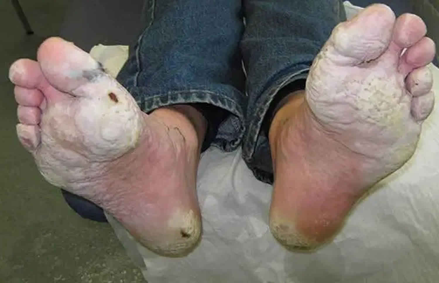
Trench foot often begins with a tingling, itching and/or numb sensation. Due to the poor blood supply, the feet may become erythematous (red) or cyanotic (bluish), and they may have an odor of decay if necrosis has started to set in. Significant swelling can occur, and there are some descriptions that the feet can double in size due to the edema 4. Trench foot disease has been known to affect just the heels or toes, but most commonly, it involves the entire foot. Some patients have described an itching or tingling sensation while others have described a prickly feeling and a heaviness to the feet. The skin can appear blotchy, and as the disease progresses, blisters and open sores can occur which can lead to fungal as well as bacterial infections. As the disease continues to advance, skin and tissue may slough off. If the condition is left untreated, gangrene can set in involving the toes, heel, or entire foot. At this point, amputation may be needed to avoid further progression of the disease and other complications such as sepsis and death.
Trench foot diagnosis
The diagnosis of trench foot is entirely clinical. Your doctor should inspect your feet carefully. Typically the diagnosis of a skin infection can be made clinically; however, lab tests and cultures may be ordered to rule out infections within the blood stream. Inflammatory markers such as a C-reactive protein (CRP) or erythrocyte sedimentation rate might prove helpful. X-ray, bone scan and/or MRI tests are also sometimes ordered if there is concern of infection deep to the skin or in the bone (osteomyelitis). Your doctor may prescribe oral antibiotics if necessary to treat a suspected infection. Some infections in the feet may require hospitalization. Frostbite and gangrene may also require hospitalization. Dead skin and tissues require debridement as they serve as a nidus for infection and continued inflammation.
Trench foot treatment
Treatment for trench foot is similar to the treatment for frostbite. Take the following steps 19:
- Thoroughly clean and dry your feet.
- Put on clean, dry socks daily.
- Treat the affected part by applying warm packs or soaking in warm water (102° to 110° F [38.9° to 43.3°C]) for approximately 5 minutes.
- When sleeping or resting, do not wear socks.
- Obtain medical assistance as soon as possible.
If you have a foot wound, your foot may be more prone to infection. Check your feet at least once a day for infections or worsening of symptoms.
Trench foot prognosis
Today, trench foot is usually identified early, and the treatment is very straightforward. Keeping the feet dry and warm is imperative. Rest and elevation of the affected foot are encouraged since this will help prevent new wounds and blisters. Nonsteroidal anti-inflammatory drugs (NSAIDs) will help with any discomfort the patient is having and will also help to alleviate the swelling. If the patient cannot take NSAIDs, then acetaminophen or aspirin will help with the pain but may not help with the swelling. According to the Center for Disease Control and Prevention (CDC), the prevention and treatment of trench foot are straightforward, and their recommendations are as follows:
- Take off socks
- Avoid wearing dirty socks to bed
- Wash the affected area right away
- Dry feet completely
- Apply heat packs to the area for up to 5 minutes
If the above measures do not help the condition, then the CDC recommends further evaluation by a clinician.
References- Proctor-Brown L, Hicks R, Colmer S, Guilfoyle D, Dallap-Schaer B, Johnson AL, Tomlinson J. Distal limb pathologic conditions in horses treated with sleeve-style digital cryotherapy (285 cases). Res. Vet. Sci. 2018 Dec;121:12-17.
- Ungley CC, Blackwood W. Peripheral vasoneuropathy after chilling. “Immersion foot and immersion hand”. Lancet (1942) 240(6216):447–51.10.1016/S0140-6736(00)58135-5
- US Army Research Institute of Environmental Medicine. Medical Aspects of Cold Weather Operations: A Handbook for Medical Officers. Report No. TN93-4. (1993).
- Anand P, Privitera R, Yiangou Y, Donatien P, Birch R, Misra P. Trench Foot or Non-Freezing Cold Injury As a Painful Vaso-Neuropathy: Clinical and Skin Biopsy Assessments. Front Neurol. 2017;8:514. Published 2017 Sep 29. doi:10.3389/fneur.2017.00514 https://www.ncbi.nlm.nih.gov/pmc/articles/PMC5626869
- Granberg PO. Freezing cold injury. Arctic Med Res (1991) 50(Suppl 6):76–9.
- Eglin, Clare & Golden, C. & Tipton, Mike. (2009). Classification of non-freezing cold injury in patients: an interim report. https://www.researchgate.net/publication/237731002_Classification_of_non-freezing_cold_injury_in_patients_an_interim_report
- Friedman NB. The reactions of tissue to cold; the pathology of frostbite, high altitude frostbite, trench foot and immersion foot. Am J Clin Pathol (1946) 16(10):634–9.10.1093/ajcp/16.10.634
- Irwin MS, Thorniley MS, Green CJ. An investigation into the aetiology of non-freezing cold injury using near infra red spectroscopy. Biochem Soc Trans (1994) 22(4):418S.10.1042/bst022418s
- Montgomery H. Experimental immersion foot. III. Oxygen utilization by muscle and nerve of the rabbit leg two hours after its prolonged exposure to water at 3 degrees C. Trans Am Clin Climatol Assoc (1954) 66:192–8.
- Flouris AD, Cheung SS. Influence of thermal balance on cold-induced vasodilation. J Appl Physiol (2009) 106(4):1264–71.10.1152/japplphysiol.91426.2008
- Arvesen A, Rosén L, Eltvik LP, Kroese A, Stranden E. Skin microcirculation in patients with sequelae from local cold injuries. Int J Microcirc Clin Exp (1994) 14(6):335–42.10.1159/000178852
- Lee JY, Bakri I, Matsuo A, Tochihara Y. Cold-induced vasodilation and vasoconstriction in the finger of tropical and temperate indigenes. J Therm Biol (2013) 38(2):70–8.10.1016/j.jtherbio.2012.11.004
- Maley MJ, Eglin CM, House JR, Tipton MJ. The effect of ethnicity on the vascular responses to cold exposure of the extremities. Eur J Appl Physiol (2014) 114(11):2369–79.10.1007/s00421-014-2962-2
- Irwin MS. Nature and mechanism of peripheral nerve damage in an experimental model of non-freezing cold injury. Ann R Coll Surg Engl (1996) 78(4):372–9.
- Geng Z, Tong X, Jia H. Reactive oxygen species (ROS) mediates non-freezing cold injury of rat sciatic nerve. Int J Clin Exp Med (2015) 8(9):15700–7.
- Jia J, Pollock M. The pathogenesis of non-freezing cold nerve injury. Observations in the rat. Brain (1997) 120(Pt 4):631–46.10.1093/brain/120.4.631
- Endrich B, Hammersen F, Messmer K. Microvascular ultrastructure in non-freezing cold injuries. Res Exp Med (Berl) (1990) 190(5):365–79.10.1007/BF00000043
- Large A, Heinbecker P. Nerve degeneration following prolonged cooling of an extremity. Ann Surg (1944) 120(5):742–9.10.1097/00000658-194411000-00006
- Trench Foot or Immersion Foot. https://www.cdc.gov/disasters/trenchfoot.html


