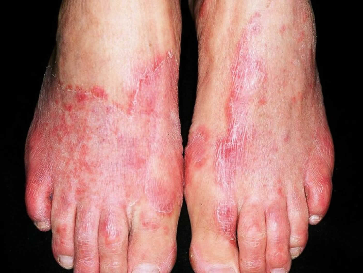
Tinea pedis
Tinea pedis also called athlete’s foot, is a superficial fungal infection of the skin of the feet caused by dermatophytes fungi, most commonly Trichophyton rubrum, Trichophyton interdigitale (previously called Trichophyton mentagrophytes var. interdigitale) and Epidermophyton floccosum 1, 2, 3. Tinea pedis dermatophytes fungi infection is usually acquired by direct contact with the causative organism, for example using a shared towel, or by walking barefoot in a public change room. The damp socks and shoes and warm, humid conditions favor the organisms’ growth. Tinea pedis commonly occurs in people whose feet have become very sweaty while confined within tight-fitting shoes. Signs and symptoms of athlete’s foot include an itchy, scaly rash. Tinea pedis or athlete’s foot is contagious and can be spread via contaminated floors, towels or clothing. People at risk of getting tinea pedis athlete’s foot infection due to occlusive footwear, sweating, and barefoot contact, include miners, soldiers, and marathon runners 4, 5, 6. Tinea pedis prevention strategies include ensuring that the spaces between your toes remain dry, wearing well-ventilated shoes and socks composed of natural fibers, and covering your feet when using communal facilities such public swimming pools or public change rooms 7, 8.
Common signs and symptoms of tinea pedis are:
- Scaly, peeling or cracked skin between the toes
- Itchiness, especially right after taking off shoes and socks
- Inflamed skin that might appear reddish, purplish or grayish, depending on your skin color
- Burning or stinging
- Blisters
- Dry, scaly skin on the bottom of the foot that extends up the side
You are at higher risk of tinea pedis or athlete’s foot if you 9:
- Wear occlusive footwear (for example, heavy industrial boots)
- Share mats, rugs, bed linens, clothes or shoes with someone who has a fungal infection
- Walk barefoot in public areas where the infection can spread, such as locker rooms, saunas, swimming pools, communal baths and showers
- Excessive sweating (hyperhidrosis)
- Live in a hot and humid environment
- Have prolonged exposure to water
- Have underlying immunodeficiency or diabetes
- Use systemic corticosteroids or immune suppressive medications
- Have poor peripheral circulation or lymphedema.
Despite the advent of anti-fungal medication, the incidence of tinea pedis has increased in recent years 10. Tinea pedis is the most common dermatophyte infection and is common worldwide, with more than 70% of the population experiencing this infection during their lifetime particularly in hot, tropical, urban environments 11. Tinea pedis onychomycosis is more prevalent in older people 12. Tinea pedis is caused by the same type of fungi (dermatophytes) that cause ringworm and jock itch. Tinea pedis may be accompanied by tinea cruris (jock itch), tinea manuum (tinea affecting the hand) or tinea unguium (fungal nail infection). Tinea pedis is often mistakenly diagnosed as candidiasis, atopic eczema, contact dermatitis, psoriasis and erythrasma 13. Most importantly, tinea pedis increases the risk of diabetic foot syndrome 14.
Tinea pedis athlete’s foot diagnosis is confirmed by the detection of segmented hyphae in skin scrapings using a potassium hydroxide (KOH) preparation and fungal culture from these skin flakes 15. Increasingly rapid real-time diagnostic polymerase chain reaction (PCR) is being used, which not only allows the rapid identification of infection and the species, but also quantification, which can prove helpful in cases of recurrence of infection where there is clinical uncertainty over resolving pre-existing infection or recurrence 16.
If you have tinea pedis on your feet, you can use an over-the-counter antifungal medicine. This includes antifungal lotion, cream, or powder. Your doctor will want you to apply it for 2 to 4 weeks. This may vary based on how bad your symptoms are or if the tinea pedis has spread.
If your tinea pedis or athlete’s foot doesn’t respond to nonprescription antifungal products and self-care, you may need to see a doctor to get a prescription-strength antifungal cream or ointment, such as clotrimazole (Lotrisone), econazole (Ecoza, Spectazole) or ciclopirox (Loprox, Penlac). If you have a more serious infection, your doctor might prescribe antifungal pills, such as terbinafine (Lamisil) or itraconazole (Sporanox, Tolsura). Or you might need both topical and oral antifungal medicine.
Figure 1. Tinea pedis
If you have a rash on your foot that doesn’t improve within two weeks of beginning self-treatment with an over-the-counter antifungal product, see your doctor.
If you have diabetes, see your doctor if you suspect that you have athlete’s foot. Also see your doctor if you have signs of an infection — swelling of the affected area, pus, fever.
Is tinea pedis contagious?
Yes. Tinea pedis or athlete’s foot is contagious and can spread through contact with an infected person or from contact with contaminated surfaces, such as towels, floors and shoes. You can also spread it from the foot to other parts of your body, especially if you scratch or pick the infected parts of your foot. It’s common for the infection to spread from your feet to your groin because the fungus can travel on hands or towels.
Who gets tinea pedis?
Tinea pedis or athlete’s foot usually occurs in males and adolescents or young adults, but can also affect females, children and older people. The mean age of onset was 15 years in one study 17. Tinea pedis or athlete’s foot is contagious and can spread through contact with an infected person or from contact with contaminated surfaces, such as towels, floors and shoes. It’s common for the infection to spread from your feet to your groin because the fungus can travel on hands or towels.
Some conditions make athlete’s foot more likely to occur:
- Living in warm, humid climates
- Using public or community pools or showers
- Wearing tight, non-ventilated footwear
- Sweating profusely
- Having diabetes or a weak immune system
Types of tinea pedis
There are 4 clinical subtypes of tinea pedis 13:
- Interdigital tinea pedis
- Moccasin tinea pedis
- Acute ulcerative tinea pedis
- Vesiculobullous (inflammatory) tinea pedis.
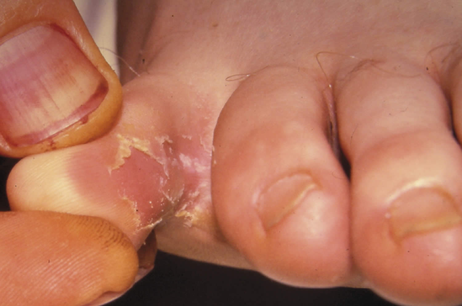
Interdigital tinea pedis
Interdigital tinea pedis is the most common form of tinea pedis. Interdigital tinea pedis typically occurs in the fourth and fifth toe web spaces but can spread to all the toe webs. Interdigital tinea pedis is characterized clinically by maceration, scaling and itching of the skin 18. Interdigital tinea pedis is caused primarily by Trichophyton interdigitale. It is often the clinical model chosen to investigate the efficacy of topical therapies in part because the thinnest skin on the foot makes the shortest treatment duration. Published efficacy rates and recommended durations of therapy for interdigital tinea pedis may not necessarily apply to other clinical forms of tinea pedis 19.
Figure 2. Interdigital tinea pedis
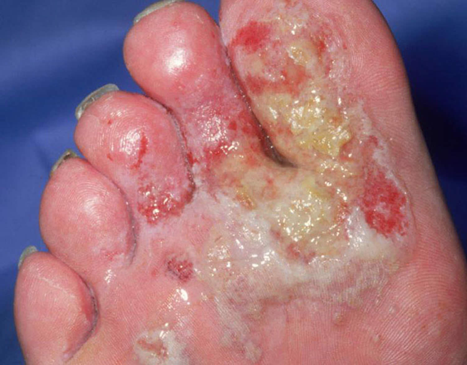
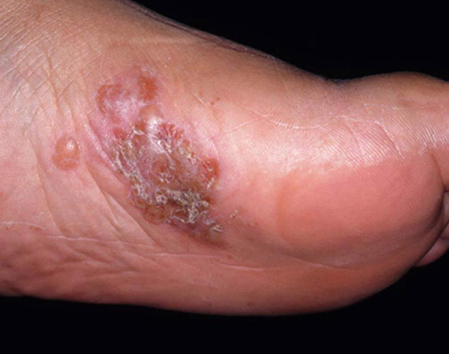
Acute ulcerative tinea pedis
Acute ulcerative tinea pedis is a variant of interdigital tinea pedis caused by Trichophyton mentagrophytes var. interdigitale, has a more aggressive clinical presentation 13, 20. Ulcerative tinea pedis is secondarily infected with Gram negative bacteria and is also known as foot intertrigo or dermatophytosis complex 21. Pseudomonas aeruginosa is isolated most frequently. This is followed in frequency by Enterococcus coli and Proteus mirabilis. Gram positive cocci (Staphylococcus and Streptococcus) are less frequent agents 22.
Acute ulcerative tinea pedis typically begins in the 3rd and 4th interdigital spaces and extends to the lateral dorsum and/or the plantar surface of the arch. These toe web lesions are usually macerated and have scaling borders and foul odor arising from the process of tissue breakdown 20, 13. Secondary bacterial infection, cellulitis, and lymphangitis are common complications.
A localized infection may be treated with a topical antibacterial such as 5% amikacin; a widespread infection can be treated with oral antibiotics with Gram negative coverage such as flucloxacillin or ciprofloxacin 22 and astringent foot soaks, rest, elevation and even hospitalization may be necessary 19.
If the lesion is exudative, a povidone-iodine solution can be useful for its antiseptic and drying properties. A solution of 1:1000 or 1:10 000 potassium permagnate solution or Burrow’s solution can be used as a wet soak if blistering infections or discharging abscesses are present.
Attention to cleaning the feet and then drying them, especially the interdigital spaces, is recommended. A gentle antiperspirant or astringent may be beneficial in people prone to excessive sweating or who need to wear occlusive footwear. Patients with diabetic neuropathy should be encouraged to inspect their feet daily to identify any skin damage.
Figure 3. Acute ulcerative tinea pedis
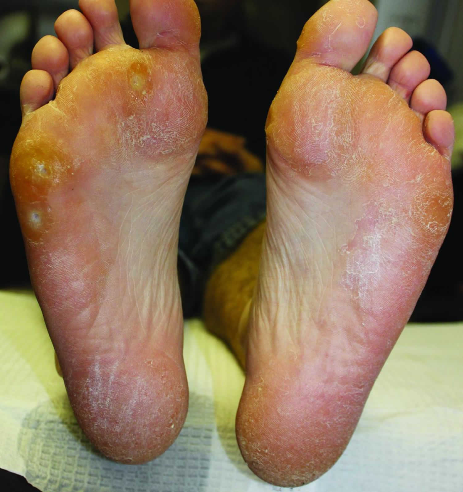
Moccasin tinea pedis
The moccasin tinea pedis or chronic dry form of tinea pedis is typically the result of Trichophyton rubrum, a keratophilic fungus 19. Moccasin tinea pedis presents as a non-inflammatory infection manifesting as mild redness (erythema) and dry silvery scales that favors the thicker keratin of the plantar aspect of the feet. Interestingly, Trichophyton rubrum actually produces penicillin in vitro and in vivo to help the infection compete with bacteria on the skin 23. The dry hyperkeratotic form of tinea pedis is concentrated within the callused areas of the sole and extends laterally to Wallace’s line, where thin dorsal skin meets the thick skin of the sole.
When the infection encompasses the entire sole, it can be called a moccasin pattern 24. The body responds to the infection with accelerated keratinization in an attempt to shed the infected keratinocytes. This hyperkeratosis may simply appear as dry skin and cracked heels. Many patients attempt to reduce this scale by using abrasive files to scrape the dead skin away, not realizing they may be spreading a fungal infection. Aggressive scraping can trigger lichen simplex chronicus and neurodermatitis.
Figure 4. Moccasin tinea pedis
Vesiculobullous tinea pedis
Trichophyton mentagrophytes is the most frequent cause of vesiculobullous tinea pedis or acute inflammatory tinea pedis. Vesiculobullous tinea pedis is characterized by tiny pruritic vesicles, pustules and multilocular bullae. Underlying erythema may be evident. The medial foot is often affected. Researchers have shown that younger active patients are more prone to infection with Trichophyton mentagrophytes 13. Trichophyton mentagrophytes is more capable of attaching to moist abraded keratin and penetrating deeper than Trichophyton rubrum, triggering a greater inflammatory host response. Trichophyton mentagrophytes is zoonotic, meaning it can spread from animals to humans through direct contact and is typically present with tinea pedis infections spread from companion animals 25.
Vesiculobullous tinea pedis develop with erythematous bases on the arch, interdigital spaces and other non-weightbearing surfaces 18. Vesiculobullous tinea pedis may start as a superficial dry scaling of the sole that subsequently drives deeper into the skin, stimulating acute inflammation and vesicle formation. Many polymorphonuclear cells accumulate, creating pustules and blisters. Symptoms of severe itching and excoriation are typical. These lesions can also become secondarily infected with Staphylococcus aureus or Streptococcus pyogenes 13.
Figure 5. Vesiculobullous tinea pedis
Tinea pedis causes
Tinea pedis is caused by dermatophytes fungi Trichophyton rubrum, Trichophyton mentagrophyte and Epidermophyton floccosum 1, 2, 3. Trichophyton rubrum accounts for about 70% of tinea pedis cases 26, 27. Tinea pedis dermatophytes fungi infection is usually acquired by direct contact with the causative organism, for example using a shared towel, or by walking barefoot in a public change room. The damp socks and shoes and warm, humid conditions favor the organisms’ growth.
Risk factors for getting tinea pedis
You are at higher risk of tinea pedis or athlete’s foot if you 9:
- Wear occlusive footwear (for example, heavy industrial boots)
- Share mats, rugs, bed linens, clothes or shoes with someone who has a fungal infection
- Walk barefoot in public areas where the infection can spread, such as locker rooms, saunas, swimming pools, communal baths and showers
- Excessive sweating (hyperhidrosis)
- Underlying immunodeficiency or diabetes
- Systemic corticosteroids or immune suppressive medications
- Poor peripheral circulation or lymphedema.
Tinea pedis prevention
There are certain things that can increase your risk of getting tinea pedis. These tips can help you avoid tinea pedis or athlete’s foot or avoid spreading it to others:
- Let your feet air out. When you can, wear sandals to let your feet air out as much as possible.
- Keep your skin as clean and dry as possible. Wash your feet daily. Use warm, soapy water and rinse and dry your feet thoroughly, especially between the toes. Apply a medicated foot powder (Tinactin, Gold Bond, others) or other medicated powder (Lotrimin AF, Zeasorb, others) if you’re prone to athlete’s foot.
- Change socks regularly. Change your socks at least once a day — more often if your feet get really sweaty. Moisture-wicking socks, such as those made from cotton, help keep your feet drier than do nylon socks.
- Keep your fingernails and toenails short and clean.
- Do not go barefoot in public areas, such as bathrooms, locker rooms, and showers. Wear waterproof sandals or shoes around public pools, showers and lockers rooms.
- Wear shoes that provide airflow.
- Alternate pairs of shoes. Use different shoes from day to day. This gives your shoes time to dry after each use.
- Wash your clothes regularly; don’t wear the same underwear or socks for more than one day.
- Shower right after playing a contact sport or swimming in a public pool.
- Clean household surfaces with antibacterial cleaners.
- Avoid touching people or pets that have tinea pedis. Also avoid touching the things they touch.
- Do not share items with people who have tinea pedis. If you live with others, don’t share shoes or unwashed bedding and towels.
Tinea pedis signs and symptoms
Tinea pedis or athlete’s foot may affect one or both feet. It can look different depending on which part of the foot (or feet) is involved and which fungus (ie, dermatophyte) has caused the infection:
Tinea pedis tends to be asymmetrical, and may be unilateral. It usually presents in one of three ways 9:
- Itchy erosions and/or scales between the toes, especially between 4th and 5th toes
- Scale covering the sole and sides of the feet (hyperkeratotic tinea pedis or moccasin tinea pedis, usually caused by Tinea rubrum)
- Small to medium-sized blisters, usually affecting the inner aspect of the foot (vesiculobullous type).
- On the top of the foot, athlete’s foot appears as a red scaly patch or patches, ranging in size from 1 to 5 cm. The border of the affected skin may be raised, with bumps, blisters, or scabs. Often, the center of the lesion has normal-appearing skin with a ring-shaped edge, leading to the descriptive but inaccurate name ringworm. (It is inaccurate because there is no worm involved.)
- Between the toes (the interdigital spaces), athlete’s foot may appear as inflamed, scaly, and soggy tissue. Splitting of the skin (fissures) may be present between or under the toes. This form of athlete’s foot tends to be quite itchy.
- On the sole of the foot (the plantar surface), athlete’s foot may appear as pink-to-red skin with scales ranging from mild to widespread (diffuse).
- Another type of tinea pedis infection, called bullous tinea pedis, has painful and itchy blisters on the arch (instep) and/or the ball of the foot.
- The most severe form of tinea pedis infection, called ulcerative tinea pedis, appears as painful blisters, pus-filled bumps (pustules), and shallow open sores (ulcers). These lesions are especially common between the toes but may involve the entire sole. Because of the numerous breaks in the skin, lesions commonly become infected with bacteria. Ulcerative tinea pedis occurs most frequently in people with diabetes and others with weak immune systems.
Tinea pedis can also uncommonly cause oozing and ulceration between the toes (ulcerative type), or pustules (these are more common in tinea pedis due to Tinea interdigitale than that due to Tinea rubrum).
Tinea pedis complications
The tinea pedis or athlete’s foot infection can spread to other warm, moist parts of your body. Jock itch is often caused by the same fungus that results in athlete’s foot. It’s common for the infection to spread from the feet to the groin because the fungus can travel on hands or towels.
Athlete’s foot can sometimes lead to secondary bacterial infections.
Tinea pedis diagnosis
Your doctor may be able to diagnose tinea pedis or athlete’s foot simply by looking at it, in most cases. Other typical sites, such as toenails, groin, and palms of the hands, should be examined for fungal infection, which may support a diagnosis of tinea pedis. Some types of tinea pedis or athlete’s foot look like dry skin or dermatitis. To help confirm the diagnosis and rule out other conditions, your doctor might take a skin scraping from the affected area for testing in a lab. Collection of an adequate specimen for a potassium hydroxide (KOH) preparation or fungal culture is important. In patients with tinea pedis, the highest yield may be obtained from skin scrapings taken from the active border of a plaque or patch. In vesicobullous tinea pedis, the roof of a vesicle can provide an adequate specimen. Skin scrapings are sent for microscopy in potassium hydroxide (KOH) where segmented hyphae may be observed and fungal culture from these skin flakes (mycology). Culture may not be necessary if typical fungal elements are observed on microscopy.
In general, tinea pedis is histologically characterized by acanthosis and hyperkeratosis with a sparse, superficial, perivascular infiltrate in the dermis. The vesiculobullous forms shows spongiosis and parakeratosis. Staining with periodic acid Schiff or methenamine silver stain can help to demonstrate characteristic fungal filaments 28.
Tinea pedis differential diagnosis
There are a number of other conditions that should be considered when a patient presents with suspected tinea pedis, including:
- Onychomycosis (fungal nail infection) – approximately one-third of patients with tinea pedis have a concomitant nail infection which can result in recurrent tinea pedis infections
- Dermatophytide (ide reaction or id reaction) – an allergic rash (secondary eczematisation) caused by an inflammatory fungal infection
- Non-dermatophyte associated podopompholyx – a type of eczema which can affect the feet and may resemble a vesicular form of tinea pedis. It is more likely to be bilateral and symmetrical; mycology is negative.
- Palmoplantar psoriasis.
- Palmoplantar pustulosis – typically characterised by scaly, partially or completely red, dry and thickened skin on the plantar surface of both feet, that is similar in appearance to psoriasis on other parts of the body. Note that psoriatic nail dystrophy often closely resembles onychomycosis.
- Juvenile plantar dermatosis – characterised by dry, shiny, glazed skin on the sole of the foot due primarily to friction. It is most commonly seen in children who are atopic, particularly boys aged four to eight years.
- Contact dermatitis – sweat, friction and home-remedies can cause irritant contact dermatitis; contact allergy may be due to accelerants (from rubber), chrome (leather tanning agent), glues and dyes in footwear
- Atopic dermatitis – usually diagnosed because of its presence on other body sites
- Bacterial foot infections, e.g. pitted keratolysis (associated with very malodorous feet) and bacterial web infections secondary to tinea pedis infection
Tinea pedis treatment
Once the diagnosis of tinea pedis or athlete’s foot has been confirmed, your physician will probably start treatment with an antifungal medication. Most infections can be treated with topical creams and lotions, including:
- Over-the-counter preparations such as terbinafine, clotrimazole, or miconazole
- Prescription-strength creams such as econazole, oxiconazole, ciclopirox, ketoconazole, sulconazole, naftifine, or butenafine
Other topical medications your physician may consider:
- Compounds containing urea, lactic acid, or salicylic acid to help dissolve the scale and allow the antifungal cream to penetrate better into the skin
- Solutions containing aluminum chloride, which reduces sweating of the foot
- Antibiotic creams to prevent or treat bacterial infections, if present
The safest tinea pedis treatment is topical antifungals, but recurrence is common and treatment must often be prolonged 20. Moisture reduction on the feet and in footwear is necessary for preventing recurrence. Permeable or open-toe footwear and sock changes are important especially during warm weather. Interdigital spaces should be manually dried after bathing. Drying agents are also recommended; options include antifungal powders (eg, miconazole), gentian violet, Burow solution (5% aluminum subacetate) soaks, and 20 to 25% aluminum chloride solution nightly for 1 week then 1 to 2 times/week as needed.
Rarely, more extensive infections or those not improving with topical antifungal medications may require 3–4 weeks of treatment with oral antifungal pills, including:
- Terbinafine
- Itraconazole
- Griseofulvin
- Fluconazole
- Ketoconazole
Systemic therapy is indicated in the treatment of chronic tinea pedis when repeated attempts to clear an infection with topicals have failed. Laboratory confirmation of the diagnosis is appropriate before systemic therapy begins. A careful review of both the medical and allergy history is important. If you suspect hepatic dysfunction, order appropriate laboratory tests. Short courses of systemic antifungal medication, like short courses of systemic antibiotics, generally do not require baseline hepatic function testing as long as the patient does not have a history of hepatitis or cirrhosis. Terbinafine and itraconazole should be the preferred systemic drugs. Minimum duration of treatment should be 2–4 weeks in naive cases and > 4 weeks in recalcitrant cases 29. Itraconazole 200 mg orally once a day for 1 month (or pulse therapy with 200 mg 2 times a day 1 week/month for 1 to 2 months) and terbinafine 250 mg orally once a day for 2 to 6 weeks. Concomitant topical antifungal use may reduce recurrences. The infection should go away within 4–6 weeks after using effective treatment.
Tinea pedis home remedies
These tips can help you ease the symptoms of athlete’s foot or avoid a recurrence:
- Keep your feet clean and dry. Wash your feet twice a day and gently towel-dry between the toes, even using a hair dryer (on low setting) if possible. Use a separate towel for your feet, and do not share this towel with anyone else.
- Use an antifungal product. After washing and drying your feet, apply an antifungal product. The over-the-counter antifungal terbinafine has been shown to be very effective. Another option is clotrimazole or miconazole. You may need to experiment to find the product and formulation — ointment, gel, cream, lotion, powder or spray — that work for you. Apply the product to the affected skin as directed — usually twice a day until a week after the rash clears up. It might take 2 to 4 weeks to see results. Continue to apply the antifungal cream between your toes and to the soles of your feet for at least 2 weeks after the areas are completely clear of lesions. If the condition comes back, you might need to start applying the product again.
- Wear socks made of cotton or wool, and change them once or twice a day, or even more often if they become damp.
- Change socks regularly. Change your socks at least once a day — more often if your feet get really sweaty.
- Wear light, well-ventilated footwear. Avoid shoes made of synthetic material, such as vinyl or rubber. Wear sandals when possible to let your feet air out.
- Alternate pairs of shoes. Use different shoes from day to day. This gives your shoes time to dry after each use.
- Apply antifungal powder to your feet and inside your shoes every day.
- Protect your feet in public places. Wear waterproof sandals or shoes around public pools, showers and lockers rooms.
- Try not to scratch the rash. You can try soothing your itchy feet by soaking them in cool water.
- Don’t share shoes. Sharing risks spreading a fungal infection.
Tinea pedis prognosis
Prognosis for tinea pedis is good provided preventive precautions are taken 28. Hyperhidrosis is a predisposing factor for tinea infections. Because tinea pedis often starts on the feet, you should dry your toes thoroughly after bathing. Dryness of your feet including the spaces between your toes is essential if reinfection is to be avoided. The use of a good antifungal powder on your feet after bathing, particularly between the toes, is strongly advised for keeping the part dry in susceptible persons. Tolnaftate or clotrimazole powder is an excellent antifungal dusting powder for the feet. Plain talc may be dusted onto socks and shoes to keep the feet dry. Periodic use of a topical antifungal agent may be required, especially when hot occlusive footwear is worn. You may need to experiment to find the product and formulation — ointment, gel, cream, lotion, powder or spray — that work for you.
- Hsu A.R., Hsu J.W. Topical Review: Skin Infections in the Foot and Ankle Patient. Foot Ankle Int. 2012;33:612–619. doi: 10.3113/FAI.2012.0612[↩][↩]
- Rinaldi M.G. Dermatophytosis: Epidemiological and microbiological update. J. Am. Acad. Dermatol. 2000;43:S120–S124. doi: 10.1067/mjd.2000.110378[↩][↩]
- Ward H, Parkes N, Smith C, Kluzek S, Pearson R. Consensus for the Treatment of Tinea Pedis: A Systematic Review of Randomised Controlled Trials. J Fungi (Basel). 2022 Mar 29;8(4):351. doi: 10.3390/jof8040351[↩][↩]
- Macura A.B. Dermatophyte infections. Int. J. Dermatol. 1993;32:313–323. doi: 10.1111/j.1365-4362.1993.tb01464.x[↩]
- Pickup T.L., Adams B.B. Prevalence of Tinea Pedis in Professional and College Soccer Players versus Non-Athletes. Clin. J. Sport Med. 2007;17:52–54. doi: 10.1097/JSM.0b013e31802ed88e[↩]
- Caputo R., De Boulle K., Del Rosso J., Nowicki R. Prevalence of superficial fungal infections among sports-active individuals: Results from the Achilles survey, a review of the literature. J. Eur. Acad. Dermatol. Venereol. 2001;15:312–316. doi: 10.1046/j.1468-3083.2001.00289.x[↩]
- Gupta A.K., Cooper E.A. Update in Antifungal Therapy of Dermatophytosis. Mycopathol. Mycol. Appl. 2008;166:353–367. doi: 10.1007/s11046-008-9109-0[↩]
- Crawford F., Hollis S. Topical treatments for fungal infections of the skin and nails of the foot. Cochrane Database Syst. Rev. 2007;2007:CD001434. doi: 10.1002/14651858.CD001434.pub2[↩]
- Tinea pedis. https://dermnetnz.org/topics/tinea-pedis[↩][↩][↩]
- Havlickova, B., Czaika, V.A. and Friedrich, M. (2008), Epidemiological trends in skin mycoses worldwide. Mycoses, 51: 2-15. https://doi.org/10.1111/j.1439-0507.2008.01606.x[↩]
- Brooks KE, Bender JF. Tinea pedis: diagnosis and treatment. Clin Podiatr Med Surg. 1996 Jan;13(1):31-46.[↩]
- Kromer C., Celis D., Hipler U., Zampeli V.A., Mößner R., Lippert U. Dermatophyte infections in children compared to adults in Germany: A retrospective multicenter study in Germany. JDDG J. Dtsch. Dermatol. Ges. 2021;19:993–1001. doi: 10.1111/ddg.14432[↩]
- Hsu AR, Hsu JW. Topical review: skin infections in the foot and ankle patient. Foot Ankle Int. 2012 Jul;33(7):612-9. https://doi.org/10.3113/FAI.2012.0612[↩][↩][↩][↩][↩][↩]
- Eckhard, M., Lengler, A., Liersch, J., Bretzel, R.G. and Mayser, P. (2007), Fungal foot infections in patients with diabetes mellitus – results of two independent investigations. Mycoses, 50: 14-19. https://doi.org/10.1111/j.1439-0507.2007.01425.x[↩]
- Levitt J.O., Levitt B.H., Akhavan A., Yanofsky H. The Sensitivity and Specificity of Potassium Hydroxide Smear and Fungal Culture Relative to Clinical Assessment in the Evaluation of Tinea Pedis: A Pooled Analysis. Dermatol. Res. Pract. 2010;2010:764843. doi: 10.1155/2010/764843[↩]
- Miyajima Y., Satoh K., Uchida T., Yamada T., Abe M., Watanabe S.-I., Makimura M., Makimura K. Rapid real-time diagnostic PCR for Trichophyton rubrum and Trichophyton mentagrophytes in patients with tinea unguium and tinea pedis using specific fluorescent probes. J. Dermatol. Sci. 2013;69:229–235. doi: 10.1016/j.jdermsci.2012.11.589[↩]
- Mazza M, Refojo N, Davel G, Lima N, Dias N, Passos da Silva CMF, Canteros CE; Mycology Network of the Province of Buenos Aires (MNPBA). Epidemiology of dermatophytoses in 31 municipalities of the province of Buenos Aires, Argentina: A 6-year study. Rev Iberoam Micol. 2018 Apr-Jun;35(2):97-102. doi: 10.1016/j.riam.2017.07.002[↩]
- Joseph WS. A closer look at topicals for tinea pedis. Podiatry Today. 2009; 22(9):48–56. https://www.hmpgloballearningnetwork.com/site/podiatry/a-closer-look-at-topicals-for-tinea-pedis[↩][↩]
- Treatment Principles For Tinea Pedis. https://www.hmpgloballearningnetwork.com/site/podiatry/treatment-principles-tinea-pedis[↩][↩][↩]
- Tinea Pedis (Athlete’s Foot). https://www.msdmanuals.com/professional/dermatologic-disorders/fungal-skin-infections/tinea-pedis-athlete-s-foot[↩][↩][↩]
- Dermatophyte (tinea) infections. https://www.uptodate.com/contents/dermatophyte-tinea-infections[↩]
- Aste N, Atzori L, Zucca M, Pau M, Biggio P. Gram-negative bacterial toe web infection: a survey of 123 cases from the district of Cagliari, Italy. J Am Acad Dermatol. 2001 Oct;45(4):537-41. doi: 10.1067/mjd.2001.114747[↩][↩]
- Youssef N, Wyborn CH, Holt G. Antibiotic production by dermatophyte fungi. J Gen Microbiol. 1978 Mar;105(1):105-11. doi: 10.1099/00221287-105-1-105[↩]
- James WD, Elston DM, Berger TG, Andrews GC. Andrews’ Diseases of the Skin: Clinical Dermatology. Saunders/Elsevier, London, 2011, pp. 293-294.[↩]
- Hygiene-related Diseases. https://www.cdc.gov/healthywater/hygiene/disease/athletes_foot.html[↩]
- Lipner SR, Scher RK. Onychomycosis: Clinical overview and diagnosis. J Am Acad Dermatol. 2019 Apr;80(4):835-851. doi: 10.1016/j.jaad.2018.03.062[↩]
- R. Wang, Y. Song, M. Du, E. Yang, J. Yu, Z. Wan, R. Li, Skin microbiome changes in patients with interdigital tinea pedis, British Journal of Dermatology, Volume 179, Issue 4, 1 October 2018, Pages 965–968, https://doi.org/10.1111/bjd.16706[↩]
- Nigam PK, Saleh D. Tinea Pedis. [Updated 2022 Jul 3]. In: StatPearls [Internet]. Treasure Island (FL): StatPearls Publishing; 2022 Jan-. Available from: https://www.ncbi.nlm.nih.gov/books/NBK470421[↩][↩]
- Rajagopalan M, Inamadar A, Mittal A, Miskeen AK, Srinivas CR, Sardana K, Godse K, Patel K, Rengasamy M, Rudramurthy S, Dogra S. Expert Consensus on The Management of Dermatophytosis in India (ECTODERM India). BMC Dermatol. 2018 Jul 24;18(1):6. doi: 10.1186/s12895-018-0073-1[↩]


